4,068 posters, 219 videos, 101 topics, 69 sessions, 14,365 authors, 6,128 institutions
ePostersLive by SciGen Technologies S.A. All rights reserved.
Alzheimer's Association International Conference 2026
July 12-15, 2026 | London, United Kingdom, and Online

9472
Regional Tau Thresholds That Mark Transitions Across The Alzheimer’s Disease Continuum
Poster Presenter
Authors
Affiliations
Florey Institute of Neuroscience and Mental Health, The University of Melbourne, Parkville, VIC 3052, Australia;, Department of Molecular Imaging & Therapy, Austin Health, Heidelberg, VIC 3084, Australia, The Australian e-Health Research Centre, CSIRO, Parkville, VIC 3052, Australia
Part of Topic
Background:
Clinical progression along the Alzheimer’s disease (AD) continuum is a key secondary endpoint in anti‑amyloid and anti‑tau therapeutic trials. However, the regional tau burden required to drive transitions between clinical stages remains poorly defined.
Methods:
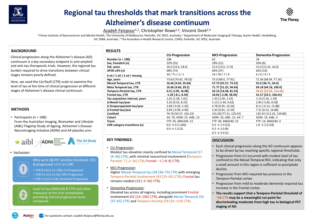
We included 188 Aβ‑positive participants (Centiloid >25) from AIBL, ADNI, and A4 who demonstrated clinical progression, defined as an increase in Clinical Dementia Rating (CDR). Tau PET (18F-Flortaucipir or 18F-MK-6240) was quantified on CenTauR (CTR) scale, in three regions of interest (ROIs): Mesial Temporal, Temporo-Parietal, and Frontal ROIs. We report the regional tau levels observed at the timepoint preceding clinical progression from cognitively unimpaired (CU), mild cognitive impairment (MCI), and mild dementia.
Results:
Among participants who progressed from the CU stage (CDR 0→≥0.5, n=108) tau elevation was observed primarily within the Mesial Temporal ROI, although levels were generally modest (median [IQR] CTR: 17 [4–36]). Tau in neocortical regions remained minimal (Temporo-Parietal: 7 [-2–16]) and Frontal: -1 [-8–8]).
Participants who progressed from MCI (CDR 0.5→≥1, n=62) showed notably higher Mesial Temporal tau (58 [36–73]), and an increase in Temporo-Parietal tau (43 [15–81]) while Frontal tau remained relatively modest (18 [-3–58]).
In those progressing from mild dementia (CDR 1→≥2, n=18) all three regions demonstrated elevated tau, with prominent involvement of the Frontal ROI (42 [24–106]) alongside increases in Mesial Temporal (55 [37–65]) and Temporo-Parietal (59 [33–114]) regions.
Conclusion:
Each clinical progression along the AD continuum appears to be driven by tau reaching specific regional thresholds. Progression from CU occurred with modest level of tau confined to the Mesial Temporal ROI, indicating that only a small amount in this region is sufficient to precipitate decline. Progression from MCI required tau presence in the Temporo‑Parietal cortex, while progression from mild to moderate dementia required tau increase in the Frontal cortex. These findings reinforce the importance of regional tau topography as the principal determinant of clinical worsening, beyond tau burden. The results further support that a Temporo‑Parietal threshold of ~60 CTR may be a meaningful cut‑point for discriminating moderate from high tau in biological PET staging of AD.
Comments
Email is required but is not shown publicly.
Loading comments...