4,068 posters, 219 videos, 101 topics, 69 sessions, 14,365 authors, 6,128 institutions
ePostersLive by SciGen Technologies S.A. All rights reserved.
Alzheimer's Association International Conference 2026
July 12-15, 2026 | London, United Kingdom, and Online

1192
Predicting Onset Of Alzheimer Disease Symptoms With The Interaction Of Plasma %p-tau217 And Age Combined With Brain Volumes
Poster Presenter
Part of Topic
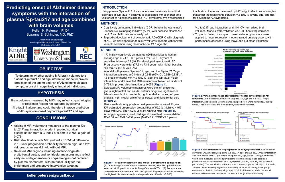
Predicting onset of Alzheimer disease symptoms with the interaction of plasma %p‑tau217 and age combined with brain volumes
Kellen K. Petersen, PhD¹; Suzanne E. Schindler, MD, PhD¹
¹Department of Neurology, Washington University School of Medicine, St. Louis, MO, USA
Objective
To determine whether adding MRI brain volumes to a plasma %p‑tau217 and age interaction model improves prediction of the timing and risk of Alzheimer’s disease (AD) symptom onset in cognitively unimpaired individuals.
Introduction
Using plasma %p‑tau217 clock models, we previously found that older age at %p‑tau217 positivity is associated with a shorter time until onset of AD symptoms. We hypothesized that brain volumes as measured by MRI might reflect co‑pathologies that affect the relationship between %p‑tau217 levels, age, and risk for developing AD symptoms.
Methods
-
Cognitively unimpaired individuals (CDR = 0) from the Alzheimer’s Disease Neuroimaging Initiative (ADNI) with baseline plasma %p‑tau217 and MRI data were analyzed.
-
To predict development of symptomatic AD (CDR > 0 with diagnosis of AD), we developed random survival forest models with forward variable selection using plasma %p‑tau217, age, the %p‑tau217*age interaction, and 114 ICV‑normalized brain volumes, with validation via 1000 bootstrap iterations.
-
To predict timing of symptom onset, selected predictors were applied to linear regression models trained on progressors, with performance assessed using leave‑one‑out cross‑validation.
-
A total of 173 initially cognitively unimpaired ADNI participants (mean age 74.5 ± 6.5 years) were followed for 8.4 ± 3.6 years; 28 (16.2%) developed symptomatic AD. Progressors were older (77.6 vs 73.9 years) and had higher baseline %p‑tau217 (6.1% vs 3.2%).
Results
-
A model including plasma %p‑tau217, age, and the %p‑tau217*age interaction achieved a C‑index of 0.689 (95% CI: 0.529–0.838).
-
A 12‑predictor model including %p‑tau217, age, the %p‑tau217*age interaction, and 9 selected MRI volumetric measures achieved a C‑index of 0.768, improving discrimination by 0.079.
-
Selected MRI measures were the left precentral gyrus, right rostral and caudal anterior cingulate, right inferior lateral ventricle, third ventricle, right cerebellar cortex, left pars orbitalis, right medial orbitofrontal cortex, and right frontal pole.
-
Risk stratification by predicted risk percentiles showed 10‑year Kaplan–Meier–estimated progression probabilities of 52.3% (high‑risk) vs 4.0% (low‑risk) with MRI, and 44.2% vs 6.4% without MRI.
-
Among progressors, predicted age at onset had cross‑validated R² = 0.66, median absolute error (MdAE) = 2.6 years, mean absolute error (MAE) = 3.2 years, and RMSE = 3.8 years.
Hypothesis
Brain volumes measured by MRI may reflect co‑pathologies or resilience factors not captured by plasma %p‑tau217 alone and could therefore improve prediction of AD symptom onset beyond %p‑tau217 and age.
Conclusions
-
Adding 9 MRI volumetric measures to a plasma %p‑tau217*age interaction model improved survival discrimination (C‑index 0.689 to 0.768).
-
MRI‑based risk stratification yielded a 13.2‑fold difference in 10‑year progression probability between high‑ and low‑risk groups compared with a 6.9‑fold difference without MRI.
-
MRI regions including anterior cingulate, orbitofrontal cortex, and ventricular measures may capture early neurodegeneration or co‑pathologies not reflected in plasma biomarkers, with potential utility for trial enrichment and preventive intervention targeting.
Comments
Email is required but is not shown publicly.
Loading comments...