4,068 posters, 219 videos, 101 topics, 69 sessions, 14,365 authors, 6,128 institutions
ePostersLive by SciGen Technologies S.A. All rights reserved.
Alzheimer's Association International Conference 2026
July 12-15, 2026 | London, United Kingdom, and Online

10125
Tox-ad: Neurometabolic Network Reorganization In Ad Mouse Models Exposed To Metal/metalloids
Poster Presenter
Authors
Leandro Ceotto Freitas Lima, Kevin Kotredes, Juliet Moncaster, Scott Persohn, Kierra Eldridge, Juan Antonio Chong Chie, Gareth Howell, Lee Goldstein, Paul Territo
Affiliations
The Jackson Laboratory, Bar Harbor, ME, USA, Indiana University School of Medicine, Indianapolis, IN, USA, Boston University Chobanian & Avedisian School of Medicine, Boston, MA, USA; Boston University Alzheimer’s Disease Research Center, Boston, MA, USA, Indiana University School of Medicine, Indianapolis, IN, USA; 5Stark Neuroscience Research Institute
Part of Topic
TOX-AD: Neurometabolic network reorganization in AD mouse models exposed to Metal/Metalloids
Leandro C Freitas-Lima1*; Kevin P Kotredes2; Juliet A Moncaster3,4; Scott Persohn1; Kierra Eldridge1; Juan Antonio K. Chong Chie1; Gareth R Howell2; Lee E Goldstein3,4; Paul R Territo1,5
1Indiana University School of Medicine, Indianapolis, IN, USA; 2The Jackson Laboratory, Bar Harbor, ME, USA; 3Boston University Chobanian & Avedisian School of Medicine, Boston, MA, USA; 4Boston University Alzheimer’s Disease Research Center, Boston, MA, USA; 5Stark Neuroscience Research Institute
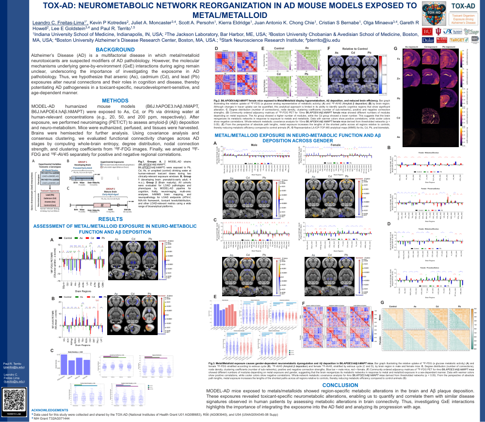
Background: Alzheimer’s Disease (AD) is a multifactorial disease in which metal/metalloid neurotoxicants are suspected modifiers of AD pathobiology. However, the molecular mechanisms underlying gene-by-environment (GxE) interactions during aging remain unclear, underscoring the importance of investigating the exposome in AD pathobiology. Thus, we hypothesize that arsenic (As), cadmium (Cd), and lead (Pb) exposures alter neural connections and their roles in cognition and disease, thereby potentiating AD pathogenesis in a toxicant-specific, neurodevelopment-sensitive, and age-dependent manner.
Methods: MODEL-AD humanized mouse models (B6J.hAPOE3.hAβ.hMAPT, B6J.hAPOE4.hAβ.hMAPT) were exposed to As, Cd, or Pb via drinking water at human-relevant concentrations (e.g., 20, 50, and 200 ppm, respectively). After exposure, we performed neuroimaging (PET/CT) to assess amyloid-β (Aβ) deposition and neuro-metabolism. Mice were euthanized, perfused, and tissues were harvested. Brains were hemisected for further analysis. Using covariance analysis and consensus clustering, we evaluated functional connectivity changes in the AD stage by calculating whole-brain entropy, degree distribution, nodal connection strength, and clustering coefficients from 18F-FDG images. Finally, we analyzed 18F-FDG and 18F-AV45 separately for positive and negative regional correlations.
Results: After exposure, these neurotoxicants altered animal behavior and accumulated in the brain, leading to aberrant Aβ deposition. Additionally, metabolic connectivity analysis revealed changes in degree distribution, connection strength, and clustering coefficients due to metal/metalloid exposure, suggesting metabolic brain network reorganization, either as compensatory mechanisms or as signs of pathological disruption. Moreover, 18F-FDG and 18F-AV45 were significantly correlated in 6/6/4/6 of 107 regions for Control/As/Cd/Pb, respectively, suggesting that Aβ deposition has a small or no significant impact on metabolic brain network reorganization.
Conclusion: MODEL-AD mice exposed to metals/metalloids showed region-specific metabolic alterations in the brain and Aβ plaque deposition. These exposures revealed toxicant-specific neurometabolic alterations, enabling us to quantify and correlate them with similar disease signatures observed in human patients by assessing metabolic alterations in brain connectivity. Thus, investigating GxE interactions highlights the importance of integrating the exposome into the AD field and of analyzing its progression over aging.
Acknowledgments: TOX-AD U01-AG088683 (LG, PT, GH); R56-AG083940 (LG, GH)
Comments
Email is required but is not shown publicly.
Loading comments...