4,068 posters, 219 videos, 101 topics, 69 sessions, 14,365 authors, 6,128 institutions
ePostersLive by SciGen Technologies S.A. All rights reserved.
Alzheimer's Association International Conference 2026
July 12-15, 2026 | London, United Kingdom, and Online

4027
Venular Degeneration Without Overt Amyloid Pathology: Reassessing The Role Of Venules In Cerebral Amyloid Angiopathy
Poster Presenter
Authors
Yuan Cao, Shaneva Edwards-Harris, Johanna Rotta, Soumya Vytla, Matthew P. Frosch, Haoyang Zhu, Theodore J. Zwang, Steven M. Greenberg, Susanne van Veluw, Valentina Perosa
Affiliations
1. J. Philip Kistler Stroke Research Center, Department of Neurology, Massachusetts General Hospital, Harvard Medical School, Boston, MA, USA. 2. Department of Neurology, The First Affiliated Hospital of Zhengzhou University, Zhengzhou, China., J. Philip Kistler Stroke Research Center, Department of Neurology, Massachusetts General Hospital, Harvard Medical School, Boston, MA, USA., Neuropathology Service, C.S. Kubik Laboratory for Neuropathology, Massachusetts General Hospital, Harvard Medical School, Boston, MA, USA., Department of Neurology, Massachusetts General Hospital, Charlestown, MA, USA., 1. Department of Neurology, Massachusetts General Hospital, Charlestown, MA, USA. 2. Harvard Medical School, Boston, MA, 02115, USA., 1. J. Philip Kistler Stroke Research Center, Department of Neurology, Massachusetts General Hospital, Harvard Medical School, Boston, MA, USA. 2. UK Dementia Research Institute, University of Edinburgh, Edinburgh, UK.
Part of Topic
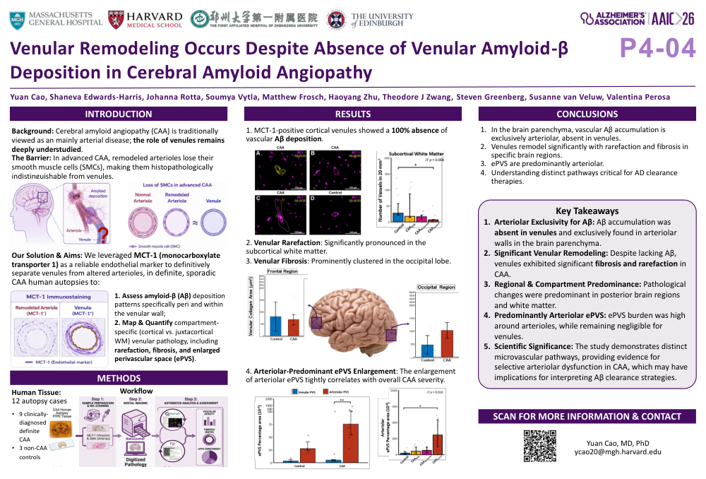
Cerebral amyloid angiopathy (CAA) is traditionally regarded as a predominantly arterial pathology, yet venular contributions remain largely unexplored. A major barrier is reliably distinguishing venules from remodeled arterioles in histopathology, because of the lack of smooth muscle cells in vessels with advanced CAA. In this study, we leveraged monocarboxylate-transporter-1 (MCT-1) as a reliable endothelial marker to differentiate venules from arterioles, enabling a comprehensive characterization of venular pathology across 12 autopsy cases (9 clinically diagnosed definite CAA and 3 non-CAA controls). Methodologically, we combined double-label immunofluorescence and 3D tissue clearing (SHIELD) with light-sheet microscopy to rigorously assess perivenular Aβ deposition. Furthermore, we employed validated semi-automated image analysis pipelines (VasOMatic and QuPath) on whole-slide images across four cortical regions to systematically quantify venular density, collagenous fibrosis, and enlarged perivascular spaces (ePVS). While we observed a complete absence of vascular amyloid-β (Aβ) deposition in MCT-1-positive cortical venules in both 2D and 3D assessments—with only rare involvement in the leptomeningeal vessels—quantitative analyses revealed significant venular rarefaction in the subcortical white matter and venular fibrosis, which was most prominent in the occipital lobe. Additionally, the percentage area of arteriolar ePVS was significantly higher than venular ePVS and increased exclusively with CAA severity. These findings confirm the historical notion that vascular Aβ accumulation in CAA is arteriolar. However, our results demonstrate that the venules are not spared and undergo structural remodeling characterized by severity-dependent rarefaction and fibrosis. Furthermore, PVS enlarge predominantly around subcortical arterioles rather than venules, possibly reflecting downstream fluid stagnation caused by upstream Aβ deposition in the arteriolar wall.
Comments
Email is required but is not shown publicly.
Loading comments...