4,068 posters, 219 videos, 101 topics, 69 sessions, 14,365 authors, 6,128 institutions
ePostersLive by SciGen Technologies S.A. All rights reserved.
Alzheimer's Association International Conference 2026
July 12-15, 2026 | London, United Kingdom, and Online

4065
Lecanemab Antibody Associates With Pre- And Postsynaptic Amyloid-ί In Human Post-mortem Alzheimer's Disease Cortex
Poster Presenter
Authors
Kristjan Holt, James Padmos, Ya Yin Chang, Giulia Albertini, Colin Smith, Jane Tulloch, Bart De Strooper, Giles Hardingham, Tara Spires-Jones
Affiliations
Institute for Neuroscience and Cardiovascular Research (INCR) and UK Dementia Research Institute (UKDRI), The University of Edinburgh, United Kingdom, Centre for Brain and Disease Research, Flanders Institute for Biotechnology (VIB), Leuven, Belgium, United Kingdom
Part of Topic
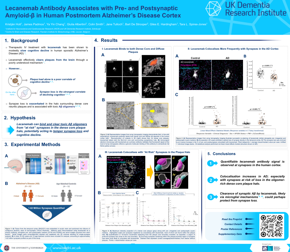
Background: Recently, lecanemab has demonstrated modest therapeutic efficacy in slowing cognitive decline for individuals diagnosed with Alzheimer’s Disease (AD)—attributed to a sustained clearance of Aβ plaques from the brain via an, as yet, poorly understood mechanism. However, it is well-understood that plaque load alone is a comparatively poor correlate of cognitive decline; thus, provoking questions as to how lecanemab acts within the brain to preserve cognition.
The strongest neuropathological correlate of cognitive decline in human AD is a region-specific loss of synapses, which is exacerbated in the halo surrounding neuritic plaques—posited to result from toxic Aβ oligomers enriched within this halo that are often seen within surviving synapses. Here, we hypothesised that, through clearing this synaptic Aβ, lecanemab could temper plaque-associated synapse loss; thereby contributing in part to slowed cognitive decline.
We aimed to investigate whether, following immunolabelling of human postmortem AD tissue with lecanemab antibody, any evidence of colocalisation with excitatory synaptic puncta could be observed. Second to this, we investigated whether excitatory synapses colocalised with lecanemab were predominantly those “at risk” within the toxic neuritic plaque halo, a region correlating spatially with profound synapse loss.
Methods: Employing array tomography (AT) for single-synapse quantification, we co-stained lecanemab with presynapses (synaptophysin) and excitatory postsynapses (PSD-95). Comparing between Braak VI AD cases (N = 20) and age-matched controls (N = 19), we performed 3-dimensional colocalisation of excitatory synapses with lecanemab in relation to disease status and/or plaque presence, profiling up to 1.62 million synapses in total.
Results: We demonstrate that lecanemab antibody positively labels extracellular plaques and exhibits increased colocalisation with excitatory synapses in AD over controls. Surviving “at risk” synapses within the synaptotoxic halo of dense core neuritic plaques more frequently colocalise with lecanemab in the AD brain.
Conclusions: Lecanemab antibody recognises Aβ within synapses and, in AD, vulnerable synapses within the neuritic plaque halo are more likely to associate with lecanemab. These data support the notion that lecanemab treatment could slow cognitive decline, at least in part, via clearance of toxic Aβ from these “at risk” synapses—or clearing dysfunctional Aβ-containing synapses themselves—likely via microglial recruitment.
Comments
Email is required but is not shown publicly.
Loading comments...