4,068 posters, 219 videos, 101 topics, 69 sessions, 14,365 authors, 6,128 institutions
ePostersLive by SciGen Technologies S.A. All rights reserved.
Alzheimer's Association International Conference 2026
July 12-15, 2026 | London, United Kingdom, and Online

2232
Lower Brain Copper Levels In Older Adults Are Linked To Less Tissue, More White Matter Hyperintensities And Enlarged Perivascular Spaces
Poster Presenter
Authors
Part of Topic
Introduction:
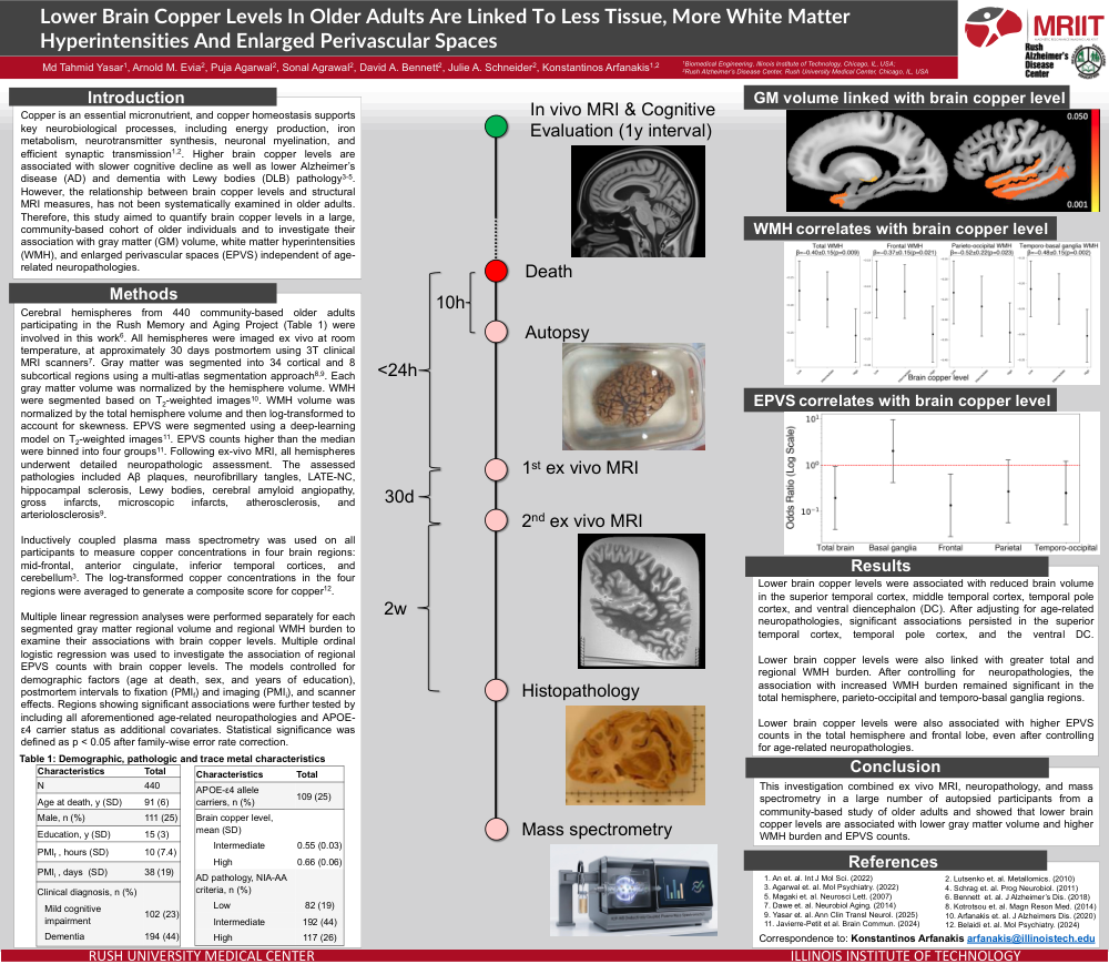
Copper is an essential micronutrient, and copper homeostasis supports key neurobiological processes, including energy production, iron metabolism, neurotransmitter synthesis, neuronal myelination, and efficient synaptic transmission. Higher brain copper levels are associated with slower cognitive decline as well as lower Alzheimer’s disease (AD) and dementia with Lewy bodies (DLB) pathology. However, the relationship between brain copper levels and structural MRI measures, has not been systematically examined in older adults. Therefore, this study aimed to quantify brain copper levels in a large, community-based cohort of older individuals and to investigate their association with gray matter (GM) volume, white matter hyperintensities (WMH), and enlarged perivascular spaces (EPVS) independent of age-related neuropathologies.
Methods:
Cerebral hemispheres from 440 community-based older adults participating in the Rush Memory and Aging Project (Table 1) were involved in this work. All hemispheres were imaged ex vivo at room temperature, at approximately 30 days postmortem using 3T clinical MRI scanners. Gray matter was segmented into 34 cortical and 8 subcortical regions using a multi-atlas segmentation approach. Each gray matter volume was normalized by the hemisphere volume. WMH were segmented based on T2-weighted images. WMH volume was normalized by the total hemisphere volume and then log-transformed to account for skewness. EPVS were segmented using a deep-learning model on T2-weighted images. EPVS counts higher than the median were binned into four groups. Following ex-vivo MRI, all hemispheres underwent detailed neuropathologic assessment. The assessed pathologies included Aβ plaques, neurofibrillary tangles, LATE-NC, hippocampal sclerosis, Lewy bodies, cerebral amyloid angiopathy, gross infarcts, microscopic infarcts, atherosclerosis, and arteriolosclerosis9.
Inductively coupled plasma mass spectrometry was used on all participants to measure copper concentrations in four brain regions: mid-frontal, anterior cingulate, inferior temporal cortices, and cerebellum. The log-transformed copper concentrations in the four regions were averaged to generate a composite score for copper.
Multiple linear regression analyses were performed separately for each segmented gray matter regional volume and regional WMH burden to examine their associations with brain copper levels. Multiple ordinal logistic regression was used to investigate the association of regional EPVS counts with brain copper levels. The models controlled for demographic factors (age at death, sex, and years of education), postmortem intervals to fixation (PMIf) and imaging (PMIi), and scanner effects. Regions showing significant associations were further tested by including all aforementioned age-related neuropathologies and APOE-ε4 carrier status as additional covariates. Statistical significance was defined as p < 0.05 after family-wise error rate correction.
Results:
Lower brain copper levels were associated with reduced brain volume in the superior temporal cortex, middle temporal cortex, temporal pole cortex, and ventral diencephalon (DC). After adjusting for age-related neuropathologies, significant associations persisted in the superior temporal cortex, temporal pole cortex, and the ventral DC.
Lower brain copper levels were also linked with greater total and regional WMH burden. After controlling for neuropathologies, the association with increased WMH burden remained significant in the total hemisphere, parieto-occipital and temporo-basal ganglia regions.
Lower brain copper levels were also associated with higher EPVS counts in the total hemisphere and frontal lobe, even after controlling for age-related neuropathologies.
Conclusion:
This investigation combined ex vivo MRI, neuropathology, and mass spectrometry in a large number of autopsied participants from a community-based study of older adults and showed that lower brain copper levels are associated with lower gray matter volume and higher WMH burden and EPVS counts.
Comments
Email is required but is not shown publicly.
Loading comments...