4,068 posters, 219 videos, 101 topics, 69 sessions, 14,365 authors, 6,128 institutions
ePostersLive by SciGen Technologies S.A. All rights reserved.
Alzheimer's Association International Conference 2026
July 12-15, 2026 | London, United Kingdom, and Online

5065
Neuropathological Correlates Of Amygdala Subregional Volumes
Poster Presenter
Authors
Amanda Annettesdotter, Katja Selezneva, Amanda E. Denning, Anika Wuestefeld, Sadhana Ravikumar, Ranjit Ittyerah, Lisa M. Levorse, Madigan Bedard, Eunice Chung, John L. Robinson, Theresa Schuck, Edward B. Lee, Sydney Lim, Winifred Trotman, Alejandra Bahena, David J. Irwin, Emilio Artacho-Pérula, María Del Mar Arroyo-Jiménez, Alicia Vela Horcajada, Esther Buendia Rosado, Noemí Villaseca González, Maria Mercedes Iñiguez De Onzoño Martin, Maria Pilar Marcos Rabal, Ana María Insausti-Serrano, Mónica Muñoz-López, David A. Wolk, Corey T. McMillan, Nicola Spotorno, Daniel T. Ohm, Ricardo Insausti, Paul A. Yushkevich, Laura E.M. Wisse
Affiliations
Lund University, Lund, Sweden, University of Pennsylvania, Philadelphia, USA, University Of Castilla-La Mancha, Albacete, Spain
Part of Topic
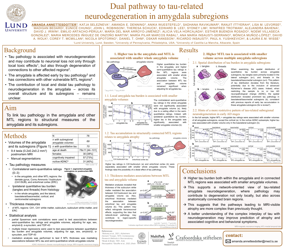
Background:The amygdala is a hotspot for neuropathologies and neurodegeneration, but whether local or distal pathology in connected regions leads to neurodegeneration and in what amygdala subregions is still unknown.We investigatedassociations between whole and subregional amygdala volumes and measures of tau, amyloid-β (Aβ), α-synuclein, and transactive response DNA-binding protein 43 (TDP-43) pathology in the amygdala and other medial temporal lobe (MTL) regions in individuals with and without neurodegenerative diseases.
Methods:Postmortem data from 78 individuals (78±11[44–101] years;38%females; 73%cognitively impaired) was included. Volumes of the whole amygdala and amygdala subregions (lateral, basolateral, basomedial, cortical, and centralmedial; for n=69 individuals) were manually segmented on 0.2x0.2x0.2 mm3 postmortemmagnetic resonance images (Fig.1). Contralateral semi-quantitativeratings of Aβ, tau, α-synuclein, and TDP-43 for the amygdala, dentate gyrus, Cornu Ammonis1/subiculum, and entorhinal cortex, and a separateipsilateral quantitative measure of amygdala tau burdenwere obtained.
Results:Higher tau ratings in MTL regions, and higher TDP-43 ratings in the amygdala and MTL, were associated with smaller whole amygdala volumes (partial Spearman correlations adjusting for age, sex, other pathologies; Fig.2a–b). The association between MTL TDP-43 and amygdala volume was not significant after adjusting for amygdala TDP-43.Average MTLand amygdalatau ratingscorrelated with lateral, basolateral, basomedial, and centralmedial subregional volumes, whereas TDP-43 was associated with all subregional volumes (Fig.2c). No associations were observed for Aβ and α-synuclein.Subregional mapping of tau pathology showed tau tangles primarily in the lateral and tau threads in the basal subregion (Fig.3a).Linear regression models (adjusting for age, sex, other pathologies) showed that higher quantitative amygdala tau burden was associated with smaller whole amygdala volume (Fig.3b), and higher lateraltau burden with smaller lateral volume (Fig.3c). No associations were found for the other subregions.
Conclusions:These findings suggest that tau pathology may contribute to amygdala neurodegenerationboth directly, through its effects within the amygdala, and indirectly through other MTL regions via retrograde transsynaptic degeneration. In contrast, TDP-43 associates to amygdala volume only directly.Subregional analyses further indicate that MTL tau and TDP-43 pathology are associated with neurodegeneration across multiple amygdala subregions.
Comments
Email is required but is not shown publicly.
Loading comments...