4,068 posters, 219 videos, 101 topics, 69 sessions, 14,365 authors, 6,128 institutions
ePostersLive by SciGen Technologies S.A. All rights reserved.
Alzheimer's Association International Conference 2026
July 12-15, 2026 | London, United Kingdom, and Online

10135
Hybrid Imaging Reveals The Molecular And Elemental Environment Of Alzheimer’S Disease In Postmortem Brain
Poster Presenter
Authors
Part of Topic
Seeking International Research Collaborators
for Disease and Anatomical Research Using Integrated Hybrid Imaging Analysis and Proteomics
Hybrid Imaging Reveals the Molecular and Elemental Environment of Alzheimer’s Disease in Postmortem Brain
Abstract
Background
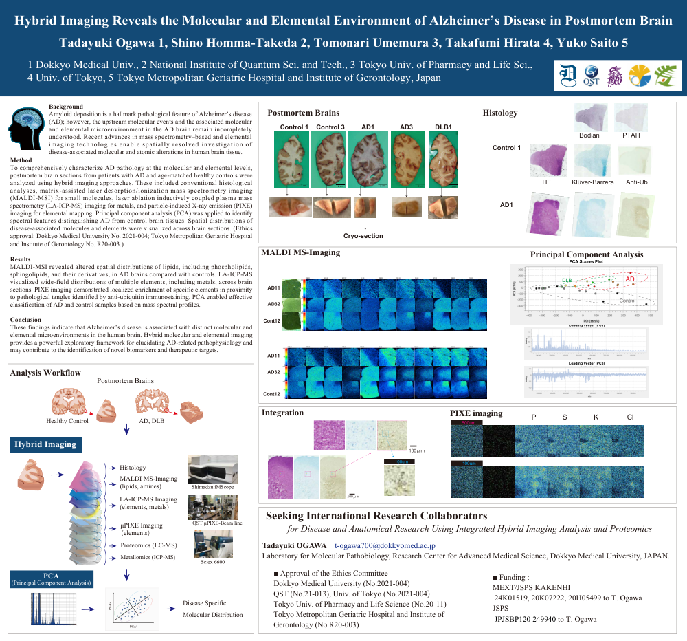
Amyloid deposition is a hallmark pathological feature of Alzheimer’s disease (AD); however, the upstream molecular events and the associated molecular and elemental microenvironment in the AD brain remain incompletely understood. Recent advances in mass spectrometry–based and elemental imaging technologies enable spatially resolved investigation of disease-associated molecular and atomic alterations in human brain tissue.
Methods
To comprehensively characterize AD pathology at the molecular and elemental levels, postmortem brain sections from patients with AD and age-matched healthy controls were analyzed using hybrid imaging approaches. These included conventional histological analyses, matrix-assisted laser desorption/ionization mass spectrometry imaging (MALDI-MSI) for small molecules, laser ablation inductively coupled plasma mass spectrometry (LA-ICP-MS) imaging for metals, and particle-induced X-ray emission (PIXE) imaging for elemental mapping. Principal component analysis (PCA) was applied to identify spectral features distinguishing AD from control brain tissues. Spatial distributions of disease-associated molecules and elements were visualized across brain sections. (Ethics approval: Dokkyo Medical University No. 2021-004; Tokyo Metropolitan Geriatric Hospital and Institute of Gerontology No. R20-003.)
Results
MALDI-MSI revealed altered spatial distributions of lipids, including phospholipids, sphingolipids, and their derivatives, in AD brains compared with controls. LA-ICP-MS visualized wide-field distributions of multiple elements, including metals, across brain sections. PIXE imaging demonstrated localized enrichment of specific elements in proximity to pathological tangles identified by anti-ubiquitin immunostaining. PCA enabled effective classification of AD and control samples based on mass spectral profiles.
Conclusion
These findings indicate that Alzheimer’s disease is associated with distinct molecular and elemental microenvironments in the human brain. Hybrid molecular and elemental imaging provides a powerful exploratory framework for elucidating AD-related pathophysiology and may contribute to the identification of novel biomarkers and therapeutic targets.
Comments
Email is required but is not shown publicly.
Loading comments...