4,068 posters, 219 videos, 101 topics, 69 sessions, 14,365 authors, 6,128 institutions
ePostersLive by SciGen Technologies S.A. All rights reserved.
Alzheimer's Association International Conference 2026
July 12-15, 2026 | London, United Kingdom, and Online

2557
Multi-ancestry Genome-wide Pleiotropy Analysis Identifies Novel Loci For Amyloid-beta Deposition And Brain Atrophy
Poster Presenter
Authors
Part of Topic
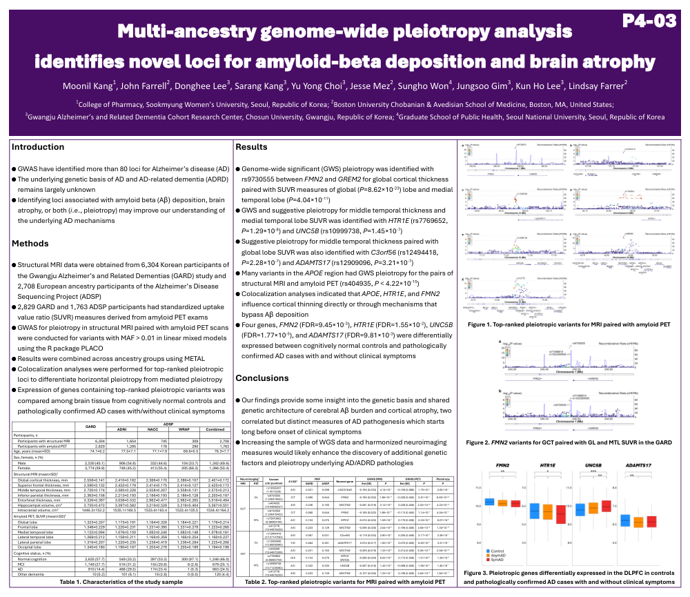
Background: Genome-wide association studies (GWAS) have identified over 80 Alzheimer’s disease (AD) loci, but the genetic basis of AD remains largely unknown. Identifying loci associated with amyloid beta (Aβ) deposition, brain atrophy, or both (i.e., pleiotropy) may improve our understanding of the underlying AD mechanisms.
Method: Structural MRI data were obtained from 6,304 Korean participants of the Gwangju Alzheimer’s and Related Dementias (GARD) Study and 2,708 European ancestry participants of the Alzheimer’s Disease Sequencing Project (ADSP), among whom 2,829 GARD and 1,763 ADSP participants also had standardized uptake value ratio (SUVR) measures derived from amyloid PET exams. GWAS for pleiotropy in paired structural MRI and amyloid PET measures were conducted for variants with MAF > 0.01 in linear mixed models using PLACO. Results were combined across ancestry groups using METAL. Colocalization analyses were performed for top-ranked pleiotropic loci to differentiate horizontal pleiotropy from mediated pleiotropy. Expression of genes containing or adjacent to top-ranked pleiotropic variants was compared among brain tissue from cognitively normal controls and pathologically confirmed AD cases with and without clinical symptoms.
Result: Genome-wide significant (GWS) pleiotropy was identified with rs9730555 between FMN2 and GREM2 for global cortical thickness paired with SUVR measures of global (P=8.62×10⁻²³) and medial temporal lobes (P=4.04×10⁻¹¹). GWS and suggestive pleiotropy for middle temporal thickness and medial temporal lobe SUVR was identified with HTR1E (rs7769652, P=1.29×10⁻⁸) and UNC5B (rs10999738, P=1.45×10⁻⁷), respectively. Suggestive pleiotropy for middle temporal thickness paired with global lobe SUVR was also identified with C3orf56 (rs12494418, P=2.28×10⁻⁷) and ADAMTS17 (rs12909096, P=3.21×10⁻⁷). Many variants in the APOE region had GWS pleiotropy for the pairs of structural MRI and amyloid PET (rs405509, P < 1.08×10⁻¹⁰). Colocalization analyses indicated that APOE, HTR1E, and FMN2 influence cortical thinning directly or through mechanisms that bypass Aβ deposition. FMN2 (FDR=9.45×10⁻³), HTR1E (FDR=1.55×10⁻²), UNC5B (FDR=1.77×10⁻⁵), and ADAMTS17 (FDR=9.81×10⁻³) were differentially expressed between cognitively normal controls and pathologically confirmed AD cases with and without clinical symptoms.
Conclusion: Our findings provide insight into the genetic basis of Aβ deposition and brain atrophy, implicated in the underlying AD mechanisms (e.g., HTR1E activates the β-arrestin pathway, involved in the pathogenesis of neurodegenerative diseases).
Comments
Email is required but is not shown publicly.
Loading comments...