4,068 posters, 219 videos, 101 topics, 69 sessions, 14,365 authors, 6,128 institutions
ePostersLive by SciGen Technologies S.A. All rights reserved.
Alzheimer's Association International Conference 2026
July 12-15, 2026 | London, United Kingdom, and Online

6993
Effects Of Physical Exercise On Brain Glucose Metabolism In A Mouse Model Of Down Syndrome: A PET Imaging Study
Part of Topic
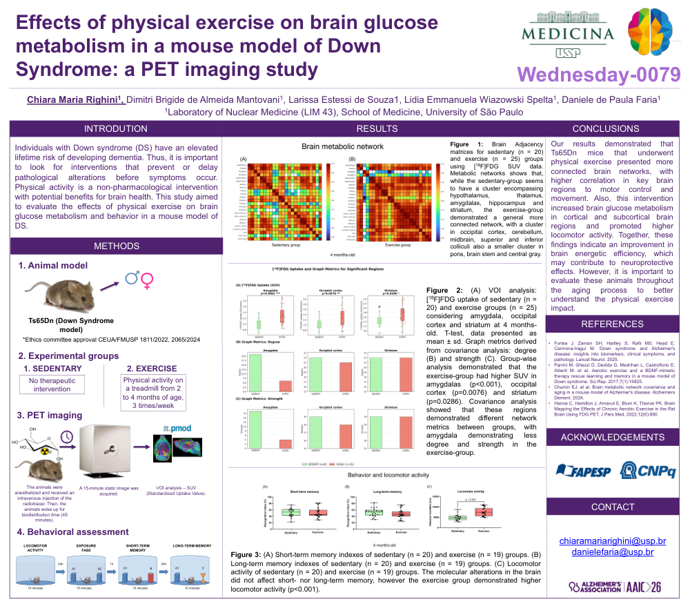
Title: Effects of physical exercise on brain glucose metabolism in a mouse model of Down Syndrome: a PET imaging study
Authors: Chiara Maria Righini, Dimitri Brigide de Almeida Mantovani, Larissa Estessi de Souza, Lidia Emmanuela Wiazowski Spelta, Daniele de Paula Faria
Background: Individuals with Down syndrome (DS) have an elevated lifetime risk of developing dementia. Thus, it is important to look for interventions that prevent or delay pathological alterations before symptoms occur. Physical activity is a non-pharmacological intervention with potential benefits for brain health. This study aimed to evaluate the effects of physical exercise on brain glucose metabolism and behavior in a mouse model of DS.
Methods: Ts65Dn trisomic mice (ethical approvals: 1811/2022;2065/2024) were divided into sedentary and exercise groups. The exercise-group performed aerobic exercise on a treadmill three times per week from 2 to 4 months of age. At 4 months, a [18F]FDG-PET acquisition using a small-animal system was performed. Images were analyzed using a volume-of-interest (VOI) approach and standardized uptake values (SUVs) were calculated and used in covariance analysis to generate metabolic networks. Behavioral assessment included locomotor activity, short- and long-term recognition memory.
Results: Group-wise analysis demonstrated that the exercise-group had higher SUV in amygdalas (p<0.001), occipital cortex (p=0.0076) and striatum (p=0.0286). Covariance analysis showed that these regions demonstrated different network metrics between groups, with amygdala demonstrating less degree and strength in the exercise-group. Metabolic networks shows that, while the sedentary-group seems to have a cluster encompassing hypothalamus, thalamus, amygdalas, hippocampus and striatum, the exercise-group demonstrated a general more connected network, with a cluster in occipital cortex, cerebellum, midbrain, superior and inferior colliculi also a smaller cluster in pons, brain stem and central gray. These molecular alterations in the brain did not affect short- nor long-term memory, however the exercise group demonstrated higher locomotor activity (p<0.001).
Conclusions: Our results demonstrated that Ts65Dn mice that underwent physical exercise presented more connected brain networks, with higher correlation in key brain regions to motor control and movement. Also, this intervention increased brain glucose metabolism in cortical and subcortical brain regions and promoted higher locomotor activity. Together, these findings indicate an improvement in brain energetic efficiency, which may contribute to neuroprotective effects. However, it is important to evaluate these animals throughout the aging process to better understand the physical exercise impact.
Comments
Email is required but is not shown publicly.
Loading comments...