4,068 posters, 219 videos, 101 topics, 69 sessions, 14,365 authors, 6,128 institutions
ePostersLive by SciGen Technologies S.A. All rights reserved.
Alzheimer's Association International Conference 2026
July 12-15, 2026 | London, United Kingdom, and Online

10138
Neural Stem Cell Extracellular Vesicles Restore Endotoxin-Induced Dysfunction In PSEN2 Human IPSC-Derived Brain Endothelial Cells
Poster Presenter
Authors
Jolene Phelps, Farnoosh Kalantarnia, Amanda Orr, Katherine Elvira, Stephanie Willerth
Affiliations
Part of Topic
Neural Stem Cell Extracellular Vesicles Restore Endotoxin-Induced Dysfunction In PSEN2 Human IPSC-Derived Brain Endothelial Cells
Background:
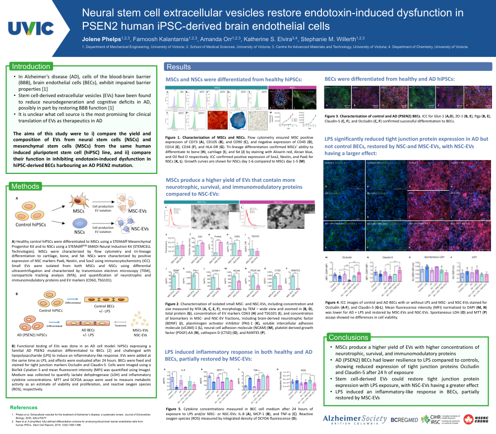
In Alzheimer’s disease (AD), brain endothelial cells (BECs) exhibit impaired barrierproperties. Stem cell-derived extracellular vesicles (EVs) have been found to reduceneurodegeneration and cognitive deficits in AD, possibly in part by restoring BBB function. EVsare lipid membrane-bound nanoparticles secreted by cells that contain bioactive cargo that canalter the phenotype of recipient cells, with stem cell-derived EVs hosting a regenerative cargo.Despite promise, it is unclear what stem cell source is the most promising for clinical translation.The aims of this study were to i) compare the yield and composition of EVs from neural stemcells (NSCs) and mesenchymal stem cells (MSCs), and ii) compare their function in inhibitingendotoxin-induced dysfunction in PSEN2 BECs.
Methods:
BECs were differentiated from human induced pluripotent stem cells (hiPSCs)expressing the PSEN2 mutation and isogenic controls using established protocols. BECs wereinduced with lipopolysaccharide (LPS) to stimulate immune response for 24 hours. EVs wereadded at the same time as LPS. To produce EVs, control hiPSCs were differentiated to NSCs andMSCs, confirmed by NSC and MSC markers and differentiation potential. EVs were isolatedusing ultracentrifugation and characterized using nanoparticle tracking analysis, transmissionelectron microscopy, EV-specific protein content, and associated neurotrophic proteinconcentrations. To evaluate function, BEC monolayers were fixed and stained for tight junctionmarkers Occludin, ZO-1, and Claudin-5. Cells were imaged and mean fluorescent intensity wasquantified using ImageJ. Medium was collected and analyzed for LDH and inflammatorycytokine concentrations.
Results:
PSEN2 and control BECs did not exhibit differences in tight junction expression, whileLPS exposure reduced expression only in PSEN2 BECs. It was found that the yield of MSC-EVswas higher and they contained higher concentrations of neurotrophic proteins. Despite this,while both NSC- and MSC-EVs restored tight junction expression during LPS exposure, NSC-EVshad a larger effect. LPS led to an increase in spontaneous LDH release only in PSEN2 BECs,inhibited by EV treatment.
Conclusion:
Reduced BEC resilience during endotoxin exposure could play a role in ADpathogenesis, with NSC-EVs representing a promising approach to mitigate harmful effects suchas reduced tight junction expression, apoptosis, and inflammation.
Comments
Email is required but is not shown publicly.
Loading comments...