4,068 posters, 219 videos, 101 topics, 69 sessions, 14,365 authors, 6,128 institutions
ePostersLive by SciGen Technologies S.A. All rights reserved.
Alzheimer's Association International Conference 2026
July 12-15, 2026 | London, United Kingdom, and Online

2169
From Hyperintensity To Edema: An Edema-specific Segmentation Algorithm On T2-flair
Poster Presenter
Authors
Hyungyou Park, Sunghwan Park, Minho Lee, Regina EY Kim, Chong Hyun Suh, Donghyeon Kim
Affiliations
Research Institute, Neurophet Inc., Seoul, Republic of Korea, Department of Radiology and Research Institute of Radiology, Asan Medical Center University of Ulsan College of Medicine, Seoul, Republic of Korea
Part of Topic
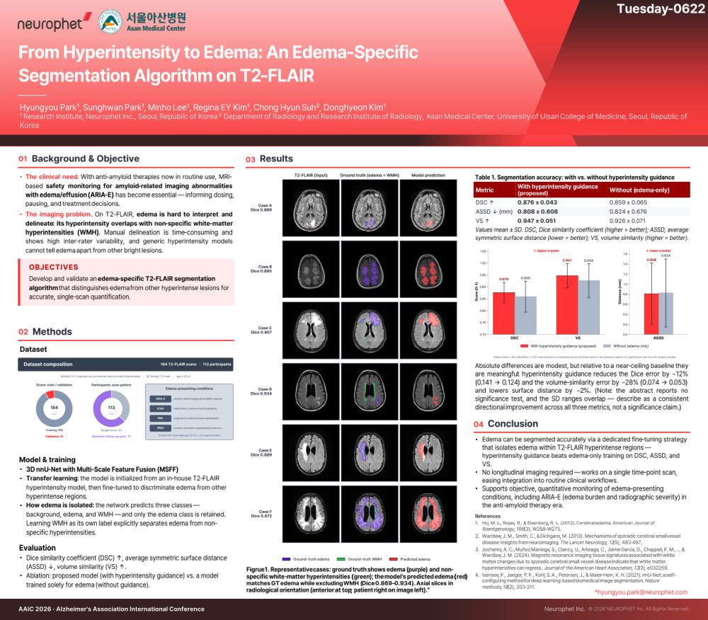
From Hyperintensity to Edema: An Edema-Specific Segmentation Algorithm on T2-FLAIR
AUTHORS
Hyungyou Park (1*), Sunghwan Park (1), Minho Lee (1), Regina EY Kim (1), Chong Hyun Suh (2), Donghyeon Kim (1)
1. Research Institute, Neurophet Inc., Seoul, Republic of Korea
2. Department of Radiology and Research Institute of Radiology, Asan Medical Center, University of Ulsan College of Medicine, Seoul, Republic of Korea
*Corresponding author: hyungyou.park@neurophet.com
Theme: Biomarkers. Topic: Neuroimaging. Subtopic: Novel imaging methods.
BACKGROUND & OBJECTIVE
The clinical need: with anti-amyloid therapies now in routine use, MRI-based safety monitoring for amyloid-related imaging abnormalities with edema/effusion (ARIA-E) has become essential, informing dosing, pausing, and treatment decisions.
The imaging problem: on T2-FLAIR, edema is hard to interpret and delineate because its hyperintensity overlaps with non-specific white-matter hyperintensities (WMH). Manual delineation is time-consuming and shows high inter-rater variability, and generic hyperintensity models cannot tell edema apart from other bright lesions.
Objective: to develop and validate an edema-specific T2-FLAIR segmentation algorithm that distinguishes edema from other hyperintense lesions for accurate, single-scan quantification.
METHODS
Dataset: 184 T2-FLAIR scans from 113 participants, sourced from multiple U.S. hospitals via a commercial data provider (Hypherdata), split into training (n = 169) and validation (n = 15) sets; 42 participants had a single scan and 71 had baseline-follow-up pairs. The dataset included edema-presenting conditions: ARIA-E, inflammatory cerebral amyloid angiopathy (iCAA), progressive multifocal leukoencephalopathy (PML), and posterior reversible encephalopathy syndrome (PRES). Ground-truth edema labels were generated by a board-certified neuroradiologist (C.H.S.) and in-house trained annotators. Cohort: 82 females / 31 males, age >= 43 years.
Model and training: a 3D nnU-Net with Multi-Scale Feature Fusion (MSFF), which captures multi-scale representations via parallel convolutions with various kernel sizes to preserve both local granularity and global context. Transfer learning: the model was initialized from an in-house T2-FLAIR hyperintensity model, then fine-tuned to discriminate edema from other hyperintense regions. How edema is isolated: the network predicts three classes - background, edema, and WMH - and only the edema class is retained; learning WMH as its own label explicitly separates edema from non-specific hyperintensities.
Evaluation: Dice similarity coefficient (DSC, higher is better), average symmetric surface distance (ASSD, lower is better), and volume similarity (VS, higher is better). Ablation: the proposed model (with hyperintensity guidance) versus a model trained solely for edema segmentation (without guidance).
RESULTS
Segmentation accuracy (with vs. without hyperintensity guidance): DSC 0.876 +/- 0.043 vs 0.859 +/- 0.065; ASSD 0.808 +/- 0.608 mm vs 0.824 +/- 0.676 mm; VS 0.947 +/- 0.051 vs 0.926 +/- 0.071 (values mean +/- SD).
Hyperintensity guidance improved every metric. Relative to a near-ceiling baseline, it reduced the Dice error by approximately 12% (0.141 to 0.124) and the volume-similarity error by approximately 28% (0.074 to 0.053), and lowered surface distance by approximately 2% - a consistent directional improvement across all three metrics (the abstract reports no significance test and the SD ranges overlap).
Qualitative: in representative cases, the model's predicted edema matched the ground-truth edema while excluding non-specific white-matter hyperintensities (Dice 0.869-0.934).
CONCLUSION
Edema can be segmented accurately via a dedicated fine-tuning strategy that isolates edema within T2-FLAIR hyperintense regions; hyperintensity guidance beats edema-only training on DSC, ASSD, and VS. The method requires no longitudinal imaging and works on a single time-point scan, easing integration into routine clinical workflows. It supports objective, quantitative monitoring of edema-presenting conditions, including ARIA-E (edema burden and radiographic severity), in the anti-amyloid therapy era.
TABLE 1 (Segmentation accuracy: with vs. without hyperintensity guidance). DSC (higher = better): 0.876 +/- 0.043 (with guidance, proposed) vs 0.859 +/- 0.065 (without, edema-only). ASSD in mm (lower = better): 0.808 +/- 0.608 vs 0.824 +/- 0.676. VS (higher = better): 0.947 +/- 0.051 vs 0.926 +/- 0.071.
FIGURE 1. Representative cases: ground truth shows edema (purple) and non-specific white-matter hyperintensities (green); the model's predicted edema (red) matches ground-truth edema while excluding WMH (Dice 0.869-0.934). Axial slices in radiological orientation (anterior at top; patient right on image left).
REFERENCES
1. Ho ML, Rojas R, Eisenberg RL. Cerebral edema. American Journal of Roentgenology. 2012;199(3):W258-W273.
2. Wardlaw JM, Smith C, Dichgans M. Mechanisms of sporadic cerebral small vessel disease: insights from neuroimaging. The Lancet Neurology. 2013;12(5):483-497.
3. Jochems ACC, Munoz Maniega S, Clancy U, et al. MRI tissue signatures associated with white matter changes due to sporadic cerebral small vessel disease indicate that white matter hyperintensities can regress. Journal of the American Heart Association. 2024;13(3):e032259.
4. Isensee F, Jaeger PF, Kohl SA, Petersen J, Maier-Hein KH. nnU-Net: a self-configuring method for deep learning-based biomedical image segmentation. Nature Methods. 2021;18(2):203-211.
KEYWORDS
edema segmentation; T2-FLAIR; ARIA-E; amyloid-related imaging abnormalities with edema; anti-amyloid therapy; MRI safety monitoring; white-matter hyperintensities; WMH; edema vs WMH discrimination; nnU-Net; Multi-Scale Feature Fusion; MSFF; transfer learning; fine-tuning; deep learning segmentation; Dice similarity coefficient; DSC; average symmetric surface distance; ASSD; volume similarity; VS; single time-point scan; inflammatory cerebral amyloid angiopathy; iCAA; progressive multifocal leukoencephalopathy; PML; posterior reversible encephalopathy syndrome; PRES; quantitative neuroimaging biomarker; brain MRI segmentation; Neurophet; Asan Medical Center.
Comments
Email is required but is not shown publicly.
Loading comments...