4,068 posters, 219 videos, 101 topics, 69 sessions, 14,365 authors, 6,128 institutions
ePostersLive by SciGen Technologies S.A. All rights reserved.
Alzheimer's Association International Conference 2026
July 12-15, 2026 | London, United Kingdom, and Online

10019
Application Of Shivai Automatic Perivascular Spaces Counting In A Memory Clinic Cohort
Poster Presenter
Authors
Carlo Tamba, Giulia Bommarito, Patrik Michel, Maëlle M. Guillemette Chavand-de-Bignicourt, Paolo Salvioni Chiabotti, Silvia Pistocchi, Yasser Aleman-Gomez, Vincent Dunet, Bianca Mazini, Olivier Rouaud, Patric Hagmann, Alessandra Griffa, Gilles Allali
Affiliations
Leenaards Memory Center, Dept. of Clinical Neurosciences, CHUV-UNIL, Stroke Center, Neurology Service, Dept. of Clinical Neurosciences, CHUV-UNIL, Dept. of Diagnostic & Interventional Radiology, CHUV-UNIL, Leenaards Memory Center, Dept. of Clinical Neurosciences, CHUV-UNIL; Neuro-X Institute, Medical Image Processing Laboratory, EPFL
Part of Topic
Application of SHIVAI Automatic Perivascular Spaces Counting in a Memory Clinic Cohort
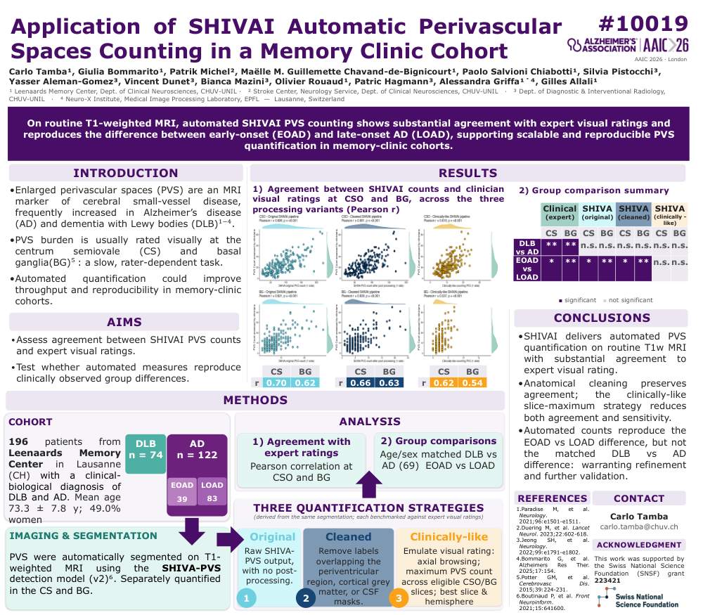
Enlarged perivascular spaces are an MRI marker of cerebral small-vessel disease and are frequently observed in neurodegenerative disorders, including Alzheimer’s disease and dementia with Lewy bodies. In clinical practice, perivascular space burden is usually assessed visually in the centrum semiovale and basal ganglia, but this approach is time-consuming and dependent on expert raters. Automated quantification may improve scalability, reproducibility, and feasibility in large memory-clinic cohorts.
In this study, we assessed the application of SHIVAI automated perivascular spaces counting in a cohort of 196 patients from the Leenaards Memory Center, CHUV Lausanne, including 74 patients with dementia with Lewy bodies and 122 patients with Alzheimer’s disease. Perivascular spaces were automatically segmented on routine T1-weighted MRI using the SHIVA-PVS detection model and quantified separately in the centrum semiovale and basal ganglia.
Three quantification strategies were compared: the original raw SHIVA-PVS output, an anatomically cleaned version excluding labels overlapping periventricular regions, cortical grey matter, or CSF masks, and a clinically-like slice-maximum strategy designed to emulate expert visual rating. Agreement between automated counts and expert visual ratings was assessed using Pearson correlations at the centrum semiovale and basal ganglia. Group comparisons were then performed to test whether automated measures could reproduce clinically observed differences, including age- and sex-matched dementia with Lewy bodies versus Alzheimer’s disease comparisons and early-onset versus late-onset Alzheimer’s disease comparisons.
Automated SHIVAI perivascular space counts showed substantial agreement with expert visual ratings, particularly when using the original and anatomically cleaned approaches. Anatomical cleaning preserved agreement with expert ratings, whereas the clinically-like slice-maximum strategy reduced both agreement and sensitivity. Automated measures reproduced the difference between early-onset and late-onset Alzheimer’s disease, supporting their ability to capture clinically meaningful variation in perivascular space burden. However, automated counts did not reproduce the matched dementia with Lewy bodies versus Alzheimer’s disease difference observed with expert visual ratings.
Overall, SHIVAI provides automated perivascular space quantification on routine T1-weighted MRI with substantial agreement to expert visual assessment. These findings support the use of automated perivascular space counting as a scalable and reproducible approach for memory-clinic cohorts, while also highlighting the need for further refinement and validation, particularly for disease-group comparisons.
Keywords: perivascular spaces; PVS; SHIVAI; SHIVA-PVS; cerebral small-vessel disease; Alzheimer’s disease; dementia with Lewy bodies; early-onset Alzheimer’s disease; late-onset Alzheimer’s disease; EOAD; LOAD; centrum semiovale; basal ganglia; T1-weighted MRI; automated MRI segmentation; memory clinic.
Comments
Email is required but is not shown publicly.
Loading comments...