4,068 posters, 219 videos, 101 topics, 69 sessions, 14,365 authors, 6,128 institutions
ePostersLive by SciGen Technologies S.A. All rights reserved.
Alzheimer's Association International Conference 2026
July 12-15, 2026 | London, United Kingdom, and Online

5048
Structural MRI-based Model For The Classification Of Predementia Stages Using Artificial Intelligence: Validation Based On CompAS Study
Poster Presenter
Authors
Xana Cid-Mejías, Sonia Valladares-Rodríguez, Miguel Ángel Rivas, Santiago Galdo-Álvarez, Mónica Lindín Novo, Cristina Lojo-Seoane, Monserrat Zurrón Ocio, Arturo X. Pereiro
Affiliations
Department of Developmental Psychology, Faculty of Psychology, Universidade de Santiago de Compostela, Santiago de Compostela, A Coruña, Spain, Department of Electronics and Computing, Higher Polytechnic Engineering School, Universidade de Santiago de Compostela, Lugo, Spain, Aging, Neuroscience and Applied Logic, Department of Psychology, Sociology and Philosophy, University of León, León, Spain, Department of Clinical Psychology and Psychobiology, Faculty of Psychology, Universidade de Santiago de Compostela, Santiago de Compostela, A Coruña, Spain
Part of Topic
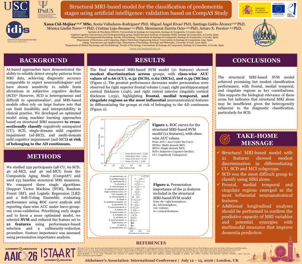
Background: AI-based approaches have demonstrated the ability of automated models to reliably detect atrophy patterns from MRI data, achieving diagnostic accuracy comparable to expert neuroradiologists (Novosad et al., 2020), and have shown sensitivity to subtle brain alterations in subjective cognitive decline (SCD) (Basanta-Torres et al., 2025). However, SCD is heterogeneous and difficult to operationalize (Röhr et al., 2020), and MRI-based models often rely on large feature sets that can limit feasibility and interpretability in clinical practice. We developed an optimized model using machine learning approaches based on structural MRI measures to classify cognitively unimpaired (CU), SCD, single-domain mild cognitive impairment (sd-MCI), and multi-domain mild cognitive impairment (md-MCI) at risk of belonging to the AD continuum.
Method: We studied 259 participants (98 CU, 69 SCD, 46 sd-MCI, and 46 md-MCI) from the Compostela Aging Study (CompAS; Juncos-Rabadán et al., 2012) and used 153 baseline structural MRI measures. We compared three single algorithms (Support Vector Machine [SVM], Random Forest [RF], and Logistic Regression [LR]) and a Soft-Voting Ensemble, evaluating performance using ROC curve analysis, and reporting class-wise AUC under leave-group-out cross-validation. Prioritizing early stages and to favor a more optimized model, we selected SVM and reduced the feature set to 21 features using performance-based selection and a collinearity-reduction procedure. Feature importance was assessed using permutation importance analysis.
Results: The final structural MRI-based SVM model (21 features) showed moderate discrimination across groups, with class-wise AUC values of 0.66 (CU), 0.52 (SCD), 0.62 (MCIs), and 0.59 (MCIm) (Figure 1). The greatest performance decreases under permutation were observed for right superior frontal volume (rh_superiorfrontal_vol; 0.049), right parahippocampal cortical thickness (rh_parahippocampal_ct; 0.046), and right rostral anterior cingulate cortical thickness (rh_rostralanteriorcingulate_ct; 0.039), highlighting frontal, medial temporal, and cingulate regions as the most influential neuroanatomical features in differentiating the groups at risk of belonging to the AD continuum (Figure 2).
Conclusion: The structural MRI-based SVM model achieved promising but modest classification performance, with frontal, medial temporal, and cingulate regions as key contributions. This supports the biological relevance of these areas but indicates that structural MRI alone may be insufficient given the heterogeneity subjacent to the diagnostic classification, particularly for SCD.
Comments
Email is required but is not shown publicly.
Loading comments...