4,068 posters, 219 videos, 101 topics, 69 sessions, 14,365 authors, 6,128 institutions
ePostersLive by SciGen Technologies S.A. All rights reserved.
Alzheimer's Association International Conference 2026
July 12-15, 2026 | London, United Kingdom, and Online

9037
Water Exchange MRI Indicates Regionally Increased Exchange And Aquaporin Activity In Alzheimer's Disease
Poster Presenter
Authors
David Alsop, Manuel Taso, Narjes Jaafar, David Shin, Joonsung Lee, Suchandrima Banerjee, Stephanie Buss, Daniel Press
Affiliations
Beth Israel Deaconess Medical Center and Harvard Medical School, Boston MA USA, 3Radiology, University of Pennsylvania, Philadelphia, PA, USA, GE HealthCare, Waukesha, WI, USA, 5GE HealthCare, Waukesha, WI, USA
Part of Topic
Introduction
Water exchange between tissue compartments reflects the water permeability of their boundaries and
their surface area. Exchange across membranes is facilitated by aquaporin channels such as AQP1 that
is highly expressed on the outer surface of the choroid plexus and AQP4 that densely lines the walls of
para-vascular spaces formed by astrocyte end feet. Reduction of AQP4 associated glymphatic transport
has been hypothesized to contribute to accumulation of amyloid in Alzheimer's Disease(AD). On the
other hand, elevated AQP4 in tissue[1] and in CSF[2] have been observed in AD. A noninvasive method to
probe aquaporin mediated exchange could provide insights into the pathology of AD and other brain
disorders.
We previously demonstrated that T2 based labeling can be used to image water exchange at the choroid
plexus and within tissue fluid boundaries of the cortex. [3] Here, we have employed a further optimized
method to evaluate changes of water exchange in patients with AD in comparison to healthy aged-
matched controls.
Methods
21 subjects with MCI or mild to moderate AD and 16 age matched healthy controls were imaged
following a protocol approved by our institutional IRB and with written consent from the subjects, and a
legally authorized representative if appropriate. Imaging was performed using the previously described
water exchange imaging on a GE Signa Premier XT 3 T scanner and the vendor's 48 channel head coil.
Imaging Method: Water exchange imaging was performed with a new approach that selectively
attenuates the magnetization in tissue, where T2 is shorter, and then observes the reduction of longer T2
signal within CSF and paravascular spaces caused by water exchange. Since water exchange across
membranes is greatly facilitated by aquaporin channels, it is expected that the resulting exchange signal
will primarily reflect aquaporin activity.
Results:
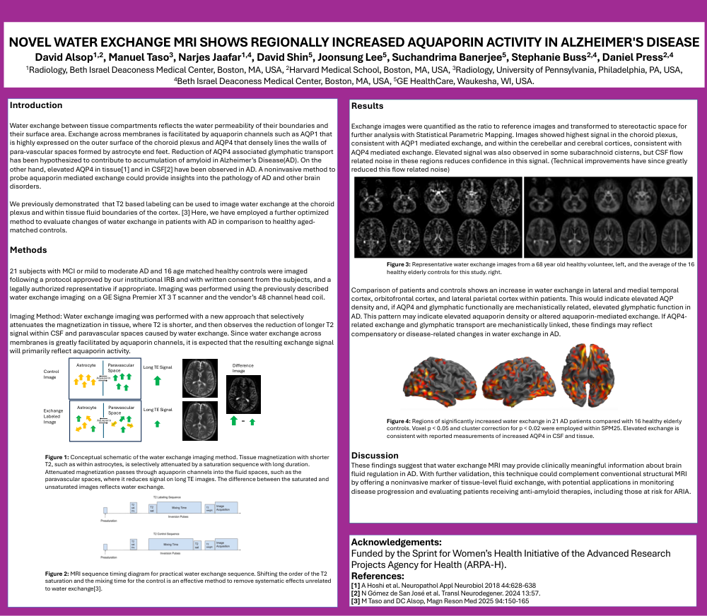
Exchange images were quantified as the ratio to reference images and transformed to stereotactic space for further analysis with Statistical Parametric Mapping. Images showed highest signal in the choroid plexus, consistent with AQP1 mediated exchange, and within the cerebellar and cerebral cortices, consistent with AQP4 mediated exchange. Elevated signal was also observed in some subarachnoid cisterns, but CSF flow related noise in these regions reduces confidence in this signal. (Technical improvements have since greatly reduced this flow related noise)
Comparison of patients and controls shows an increase in water exchange in lateral and medial temporal
cortex, orbitofrontal cortex, and lateral parietal cortex within patients. This would indicate elevated AQP
density and, if AQP4 and glymphatic functionally are mechanistically related, elevated glymphatic function in
AD. This pattern may indicate elevated aquaporin density or altered aquaporin-mediated exchange. If AQP4-
related exchange and glymphatic transport are mechanistically linked, these findings may reflect
compensatory or disease-related changes in water exchange in AD.
Acknowledgements:
Funded by the Sprint for Women's Health Initiative of the Advanced Research
Projects Agency for Health (ARPA-H).
References:
[1] A Hoshi et al. Neuropathol Appl Neurobiol 2018 44:628-638
[2] N Gómez de San José et al. Transl Neurodegener. 2024 13:57.
[3] M Taso and DC Alsop, Magn Reson Med 2025 94:150-165
Comments
Email is required but is not shown publicly.
Loading comments...