4,068 posters, 219 videos, 101 topics, 69 sessions, 14,365 authors, 6,128 institutions
ePostersLive by SciGen Technologies S.A. All rights reserved.
Alzheimer's Association International Conference 2026
July 12-15, 2026 | London, United Kingdom, and Online

10397
Towards Mapping Amyloid-Beta Plaque Load In The Human Brain With MRI At 3T
Poster Presenter
Part of Topic
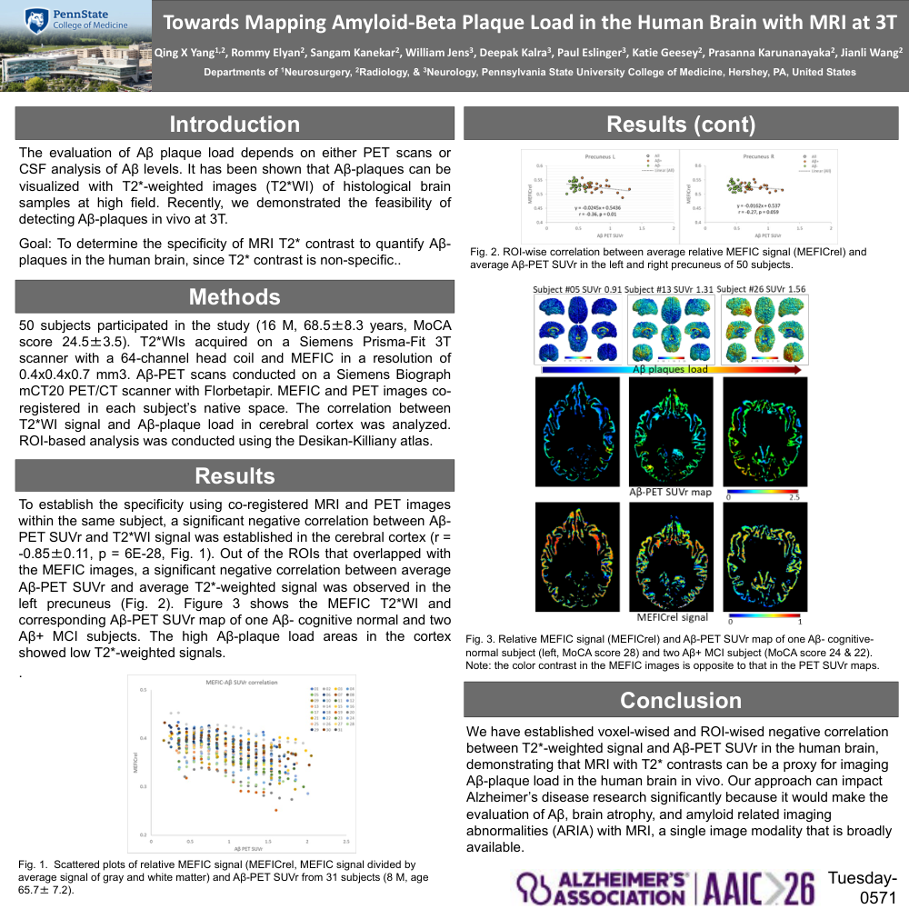
BACKGROUND: Currently the evaluation of beta-amyloid (Aβ) plaques in the human brain depends on either PET scans using radiotracers or cerebrospinal fluid analysis of Aβ level. Recently, we demonstrated the feasibility of detecting Aβ-plaques in vivo in human brain at 3T. Since T2* contrast is non-specific, the goal of this research to determine the specificity of using MRI T2* contrast to quantify Aβ-plaques load in human brain.
METHOD: Fifty subjects took part in the study (16 M, age 68.5 ± 8.3, MoCA score 24.5 ± 3.5). T2*-weighted images were acquired on a Siemens Prisma-Fit 3T scanner with a 64-channel head coil and MEFIC method in a spatial resolution of 0.4 x 0.4 x 0.7 mm3. Aβ-PET scans were conducted on a Siemens Biograph mCT20 PET/CT scanner with Florbetapir. PET and MEFIC images were co-registered in each subject’s native space. The correlation between T2*-weighted signal and Aβ-plaques load in the cerebral cortex was analyzed at individual level. ROI-based analysis was conducted using the Desikan-Killiany atlas.
RESULTS: To establish the specificity using co-registered MRI and PET images within the same subject, a significant negative correlation between Aβ-PET SUVr and T2*-weighted signal was established in the cerebral cortex (r = -0.85 ± 0.11, one-sample t-test, t = -41.1, p = 6E-28 (Figure 1). Out of the ROIs that overlapped with the MEFIC images, a significant negative correlation between average Aβ-PET SUVr and average T2*-weighted signal was observed in the left precuneus (Figure 2). Figure 3 shows the MEFIC T2*WI and corresponding Aβ-PET SUVr map of one Aβ- cognitive normal and two Aβ+ mild cognitive impairment (MCI) subjects. The high Aβ-plaque load areas in the cortex showed low T2*-weighted signals.
CONCLUSION: We have established voxel-wised and ROI-wised negative correlation between T2*-weighted signal and Aβ-PET SUVr in the human brain, demonstrating that MRI with T2* contrasts can be a proxy for imaging Aβ-plaque load in the human brain in vivo. Our approach can impact Alzheimer’s disease (AD) research significantly because it would make the evaluation of Aβ, brain atrophy, and amyloid related imaging abnormalities (ARIA) with MRI, a single image modality that is broadly available.
Comments
Email is required but is not shown publicly.
Loading comments...