4,068 posters, 219 videos, 101 topics, 69 sessions, 14,365 authors, 6,128 institutions
ePostersLive by SciGen Technologies S.A. All rights reserved.
Alzheimer's Association International Conference 2026
July 12-15, 2026 | London, United Kingdom, and Online

9126
Association Between Temporal Lobe Cortical NODDI Measures And The Plasma Biomarker GFAP
Poster Presenter
Part of Topic
BACKGROUND: Plasma glial fibrillary acidic protein (GFAP) is a blood-based biomarker of astrocytic activation that is elevated in those with Alzheimer’s disease (AD) and mild cognitive impairment (MCI) relative to cognitively unimpaired (CU) individuals. Neurite orientation dispersion and density imaging (NODDI) is a multishell diffusion MRI method capable of evaluating tissue microstructure at the axonal and dendritic level. We evaluated the associations between temporal cortex orientation dispersion index (ODI), neurite density index (NDI) and isotropic volume fraction (ISOVF) and plasma GFAP concentrations.
METHODS: 728 CU Mayo Clinic Study of Aging (MCSA) participants (ages 53-92 years) with diffusion MRIwere included. 661 individuals were cognitively unimpaired (CU), while 67 had mild cognitive impairment (MCI). NODDI imaging was obtained on a 3T Siemens Prisma scanner with 2 mm isotropicresolution. The MCALT_ADIR122 cortical atlas was used for ROI designation. Plasma testing was performedon the Fujirebio Lumipulse G1200 automated immunoassay analyzer using the Lumipulse®G GFAP Plasmakit. We fit separate linear regression models within each ROI with regional NODDI measures (neurite density index [NDI], orientation dispersion index [ODI], isotropic volume fraction [ISOVF]) as the primary predictor and log-transformed plasma GFAP concentration as the outcome. Models were adjusted for age,sex, education, structural MRI resolution, cortical thickness (or hippocampal & amygdalar volume), total intracranial volume, body mass index and amyloid PET SUVR/centiloid.
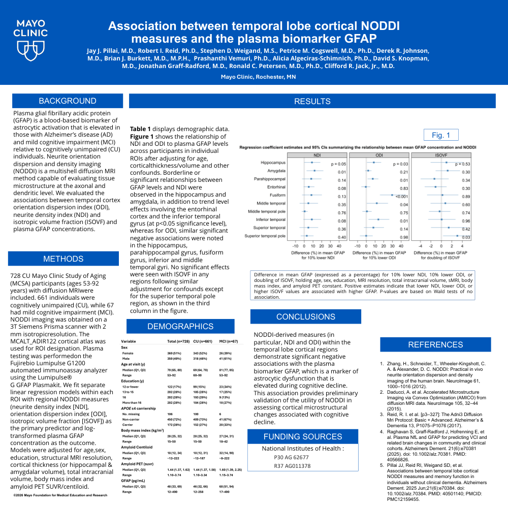
RESULTS: Table 1 displays demographic data. Figure 1 shows the relationship of NDI and ODI to plasma GFAP levels across participants in individual ROIs after adjusting for age, corticalthickness/volume and other confounds. Borderline or significant relationships between GFAP levels and NDI were observed in the hippocampus and amygdala, in addition to trend level effects involving the entorhinal cortex and the inferior temporal gyrus (at p<0.05 significance level), whereas for ODI, similar significant negative associations were noted in the hippocampus, parahippocampal gyrus, fusiform gyrus, inferior and middle temporal gyri. No significant effects were seen with ISOVF in any regions following similar adjustment for confounds except for the superior temporal pole region, as shown in the third column in the figure.
CONCLUSIONS: NODDI-derived measures (in particular, NDI and ODI) within the temporal lobe cortical regions demonstrate significant negative associations with the plasma biomarker GFAP, which is a marker of astrocytic dysfunction that is elevated during cognitive decline. This association provides preliminary validation of the utility of NODDI in assessing cortical microstructural changes associated with cognitive decline.
Comments
Email is required but is not shown publicly.
Loading comments...