4,068 posters, 219 videos, 101 topics, 69 sessions, 14,365 authors, 6,128 institutions
ePostersLive by SciGen Technologies S.A. All rights reserved.
Alzheimer's Association International Conference 2026
July 12-15, 2026 | London, United Kingdom, and Online

4311
Distinct Spatial Metabolic Patterns With Limbic Predominance Differentiate LATE From Alzheimer’s Disease
Part of Topic
Distinct spatial metabolic patterns with limbic predominance differentiate LATE from Alzheimer’s disease
Dahyun Yi, PhD, Min Soo Byun, Hyejin Ahn, Gijung Jung, Jun Sung Kim, Yoon Suk Oh, Woo Jin Cha, Yura Choi, Jun-Young Lee, Koung Mi Kang, Chul-Ho Sohn, Yun-Sang Lee, Yu Kyeong Kim, Bo Kyung Sohn, and Dong Young Lee, for the KBASE Research Group
Background
Limbic-predominant age-related TDP-43 encephalopathy (LATE) frequently co-occurs with Alzheimer’s disease (AD), yet in vivo markers that distinguish LATE-related neurodegeneration from amyloid-driven AD remain limited. Although neuropathology suggests preferential limbic involvement in LATE, it is unclear whether distinct spatial metabolic patterns beyond classification metrics are detectable in vivo, particularly in mixed pathology. We examined whether LATE is associated with an independent, limbic-predominant cerebral glucose metabolism (CM) signature that differentiates from AD.
Methods
We analyzed FDG-PET and amyloid-PET data from 520 participants (cognitively normal [CN], MCI, and AD dementia) in the Korean Brain Aging Study for the Early Diagnosis and Prediction of Alzheimer’s Disease (KBASE). Among individuals with an amnestic presentation, LATE positivity was defined by severe hippocampal atrophy and/or an abnormal inferior temporal–to–medial temporal (IT/MT) CM ratio in the absence of amyloid positivity. The IT/MT cutoff was derived from an age-adjusted normative distribution using young and old CN participants as the reference. Participants were classified into four groups: CN, LATE-only, AD-only (amyloid-positive without LATE features), and mixed LATE & AD. Voxel-wise FDG-PET analyses were performed in SPM12 to characterize group differences in spatial CM patterns. FDG SUVRs were extracted from the regions identified voxel-wise, and group differences were evaluated using ANCOVA controlling for age and sex.
Results
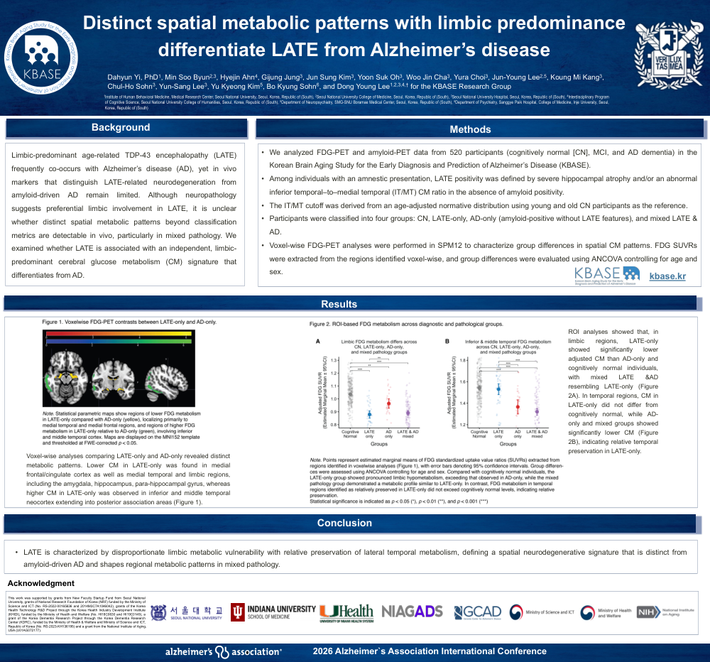
Voxel-wise analyses comparing LATE-only and AD-only revealed distinct metabolic patterns. Lower CM in LATE-only was found in medial frontal/cingulate cortex as well as medial temporal and limbic regions, including the amygdala, hippocampus, parahippocampal gyrus, whereas higher CM in LATE-only was observed in inferior and middle temporal neocortex extending into posterior association areas (Figure 1). ROI analyses showed that, in limbic regions, LATE-only showed significantly lower adjusted CM than AD-only and cognitively normal individuals, with mixed LATE &AD resembling LATE-only (Figure 2A). In temporal regions, CM in LATE-only did not differ from cognitively normal, while AD-only and mixed groups showed significantly lower CM (Figure 2B), indicating relative temporal preservation in LATE-only.
Conclusions
LATE is characterized by disproportionate limbic metabolic vulnerability with relative preservation of lateral temporal metabolism, defining a spatial neurodegenerative signature that is distinct from amyloid-driven AD and shapes regional metabolic patterns in mixed pathology.
Comments
Email is required but is not shown publicly.
Loading comments...