4,068 posters, 219 videos, 101 topics, 69 sessions, 14,365 authors, 6,128 institutions
ePostersLive by SciGen Technologies S.A. All rights reserved.
Alzheimer's Association International Conference 2026
July 12-15, 2026 | London, United Kingdom, and Online

4206
Why Some Brain Regions Rust: Identifying Biological Drivers Of Regional Brain Iron Burden In Aging
Poster Presenter
Part of Topic
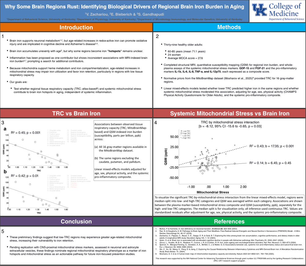
Brain iron supports neuronal metabolism, but age-related increases in redox-active iron can promote oxidative injury and are implicated in cognitive decline and Alzheimer's disease. Brain iron accumulates unevenly with age, but why some regions become iron "hotspots" remains unclear. Inflammaging has been proposed as one contributor but shows inconsistent associations with MRI-indexed brain iron burden, prompting a search for additional contributors. Because mitochondria support heme metabolism and iron compartmentalization, age-related increases in mitochondrial stress may impair iron utilization and favor iron retention, particularly in regions with low respiratory capacity. We therefore tested an atlas-based index of regional mitochondrial respiratory capacity (TRC), and systemic mitochondrial stress as contributors to iron hotspots in aging, beyond systemic inflammation.
Thirty-nine healthy older adults (ages 60-85; mean 71.1 years; 24 women) completed structural MRI, quantitative susceptibility mapping (QSM) for regional iron burden, and whole-plasma assays of the systemic mitochondrial stress markers GDF-15 and FGF-21 and the pro-inflammatory markers IL-1b, IL-6, IL-8, TNF-a, and IL-12p70, each expressed as a composite score. Normative priors from the MitoBrainMap dataset (Mosharov et al., 2025) provided TRC for 16 gray-matter regions. Linear mixed-effects models tested whether lower TRC predicted higher iron in the same regions and whether systemic mitochondrial stress moderated this association, adjusting for age, sex, physical activity, and the systemic pro-inflammatory composite.
Lower regional TRC predicted higher iron burden [R2 = 0.45; b = -63.9, p < 0.001]. A significant negative TRC by mitochondrial stress interaction [b = -8.12, 95% CI -15.6 to -0.65, p = 0.03] indicated that mitochondrial stress predicted iron burden more strongly in low-TRC regions. To visualize this effect, regions were median-split into low- and high-TRC categories and QSM was averaged within each. Systemic mitochondrial stress was positively associated with iron in low-TRC regions [R2 = 0.43, b = 17.35, p < 0.001] but not in high-TRC regions [R2 = 0.14, b = 6.45, p = 0.45].
These results suggest that low-TRC regions may experience greater age-related mitochondrial stress, increasing their vulnerability to iron retention. Pending replication with CNS-proximal mitochondrial stress markers, assessed in neuronal and astrocyte extracellular vesicles, these findings nominate regional mitochondrial respiratory phenotype as a marker of iron hotspots and mitochondrial stress as an actionable pathway for future iron-focused prevention studies.
Comments
Email is required but is not shown publicly.
Loading comments...