4,068 posters, 219 videos, 101 topics, 69 sessions, 14,365 authors, 6,128 institutions
ePostersLive by SciGen Technologies S.A. All rights reserved.
Alzheimer's Association International Conference 2026
July 12-15, 2026 | London, United Kingdom, and Online

3853
Deep Learning-Based Clinical T2-Weighted Image To Whole Brain Voxel Level Glymphatic Fluid Mapping: A Multi-Cohort Study
Part of Topic
Background:
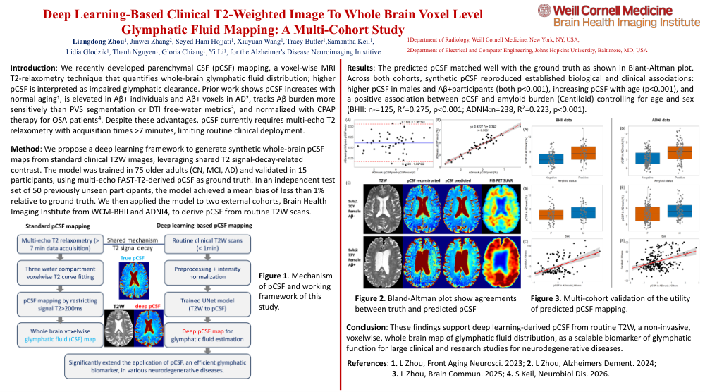
Alzheimer’s disease (AD) is increasingly viewed not only as a disorder of protein aggregationbut also of impaired neurofluid dynamics. The glymphatic system, a perivascular pathway mediating CSF-ISF exchange, supports clearance of metabolic waste, including beta-amyloid (Aβ) and tau. However, thefield lacks an efficient, scalable clinical imaging biomarker of glymphatic function, limiting mechanisticstudies and evaluation of therapies that may modulate brain clearance. We recently developedparenchymal CSF (pCSF) mapping, a voxel-wise MRI T2-relaxometry technique that quantifies whole-brainglymphatic fluid distribution; higher pCSF is interpreted as impaired glymphatic clearance. Prior workshows pCSF increases with normal aging (PMID: 37396667), is elevated in Aβ+ individuals and Aβ+ voxelsin AD (PMID: 38184796), and tracks Aβ burden more sensitively than PVS segmentation or DTI free-watermetrics (PMID: 40462860). Despite these advantages, pCSF currently requires multi-echo T2 relaxometrywith acquisition times >7 minutes, limiting routine clinical deployment.
Methods:
Here, we propose a deep learning framework to generate synthetic whole-brain pCSF mapsfrom standard clinical T2-weighted (T2W) images, leveraging shared T2 signal-decay-related contrast. Figure 1 show the mechanism. The model was trained in 75 older adults (cognitively normal, MCI, AD) andvalidated in 15 participants, using multi-echo FAST-T2-derived pCSF as ground truth. In an independenttest set of 50 previously unseen participants, the model achieved a mean bias of less than 1% relative toground truth. We then applied the model to two external cohorts—Brain Health Imaging Institute fromWeill Cornell Medicine and ADNI4—to derive pCSF from routine T2W scans.
Results:
Figure 2 show examples of predicted pCSF and Blant-Altman plot. Across both cohorts, syntheticpCSF reproduced established biological and clinical associations: higher pCSF in males and Aβ+participants (both p<0.001), increasing pCSF with age (p<0.001), and a positive association between pCSFand amyloid burden (Centiloid) controlling for age and sex (BHII: n-=125, R²=0.275, p<0.001; ADNI4:n=238, R2=0.223, p<0.001). Figure 3 shows the details.
Conclusion:
These findings support deep learning-derived pCSF from routine T2W, a non-invasive,voxelwise, whole brain map of glymphatic fluid distribution, as a scalable biomarker of glymphatic function for large clinical and research studies for neurodegenerative diseases.
Comments
Email is required but is not shown publicly.
Loading comments...