4,068 posters, 219 videos, 101 topics, 69 sessions, 14,365 authors, 6,128 institutions
ePostersLive by SciGen Technologies S.A. All rights reserved.
Alzheimer's Association International Conference 2026
July 12-15, 2026 | London, United Kingdom, and Online

9642
Innovative Retinal Microvascular Imaging Features And Cerebral Small Vessel Disease: A Systematic Review
Poster Presenter
Part of Topic
Sara Cristina Lima Rebouças1, PhD, Aicha Soumaré1, PhD, Constance Bordes1, PhD, Louis Arnould2,3, MD, PhD, Cécile Delcourt1, PhD, Jean-François Korobelnik1,4, Prof, Michel Paques5, Prof, Catherine Helmer1, MD, PhD, Stéphanie Debette6, Prof
1 University of Bordeaux, INSERM, BPH, U1219, F- 33000 Bordeaux, France.
2 Université Bourgogne Europe, CHU Dijon Bourgogne, Ophtalmologie, PEC2 UR 7460, France;
3 Ophthalmology Department, University Hospital, Dijon, France;
4 Department of Ophthalmology, Bordeaux University Hospital, F-33000 Bordeaux, France.
5 Paris Eye Imaging Group, Clinical Investigation Center 1423, Quinze-Vingts Hospital, Sorbonne Université, INSERM, Paris, France;
6 Institut du Cerveau (ICM), Paris Brain Institute, INSERM U1127, UMR CNRS 7225 Paris, Sorbonne Université, Assistance Publique des Hôpitaux de Paris, Paris, France.
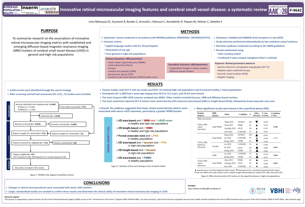
BACKGROUND: Dementia results from neurodegenerative and cerebrovascular damage, with covert cerebral small vessel disease (cSVD) representing a prevalent vascular substrate. Because retinal and cerebral microvasculatures are developmentally related and share many similarities, the retinal microvasculature may be a surrogate marker of cerebral microvasculature. Retinal microvascular changes have been associated with cSVD. Recently, innovative retinal techniques enabling in-depth assessment of retinal microvasculature emerged; these new metrics could correlate more closely with cerebral changes. This systematic review aims to summarize research on the associations of innovative retinal microvascular imaging metrics with established and emerging diffusion-based magnetic resonance imaging (MRI) markers of cSVD in general and high-risk populations.
METHODS: This work is part of a registered systematic review (PROSPERO-CRD42022327471) following PRISMA guidelines. We included English-language studies (N≥20, any age), from general or high-risk populations using retinal imaging techniques (optical coherence tomography angiography (OCT-A), adaptive optics ophthalmoscopy, dynamic vessel analyzer (DVA) or doppler). Primary outcomes comprised cross-sectional brain MRI markers (white matter hyperintensities (WMH), small subcortical infarcts, lacunes, microbleeds, perivascular spaces or composite cSVD burden). Secondary outcomes comprised longitudinal changes in these markers and diffusion-based markers. PubMed and EMBASE were searched (inception-July 2024). Study selection was performed independently in duplicate using Covidence. Narrative synthesis followed the SWiM guidelines. Results were synthetized using vote counting and/or combining P-values (wFisher’s method).
RESULTS: 6,446 records were identified. After reviewing 131 full-text reports, we included 21 studies (1,660 participants, mean ages: 44.0-72.2y, 53.6% female). Among them, 20 used OCT-A and one DVA; none analyzed other retinal imaging techniques of interest. Of the OCT-A studies, 5 examined healthy populations, 1 mixed and 14 high-risk populations. The most frequent MRI-cSVD outcome analyzed was total WMH. Other markers included lacunes, perivascular spaces, microbleeds and diffusion-based markers. The most reported OCT-A metrics included vessel density-based metrics (area or length), followed by foveal avascular zone area. The synthesis suggested that lower vessel-density-based metrics had harmful effects on cSVD markers, particularly WMH.
CONCLUSION: Changes in retinal microvasculature were associated with worst cSVD markers. Larger, standardized studies are needed to confirm these results and determine the clinical utility of innovative retinal microvascular imaging in cSVD.
Keywords: retinal microvasculature; retinal imaging; brain imaging; cerebral small vessel disease; systematic review
Comments
Email is required but is not shown publicly.
Loading comments...