4,068 posters, 219 videos, 101 topics, 69 sessions, 14,365 authors, 6,128 institutions
ePostersLive by SciGen Technologies S.A. All rights reserved.
Alzheimer's Association International Conference 2026
July 12-15, 2026 | London, United Kingdom, and Online

6151
Early And Progressive Alterations In White Matter Microstructure In 5xFAD Mice Assessed By Difussion MRI
Poster Presenter
Authors
Affiliations
Part of Topic
Regional Vulnerability of White Matter Microstructure in Early Alzheimer’s Disease: Evidence from 5xFAD Mice
A. Benítez González, V. Sasia Saldivar, H. Martínez Orozco, A.R. Aguilar Vázquez, A.J. Cisneros Mejorado, S.Y. Díaz Miranda.
Instituto de Neurobiología, Universidad Nacional Autónoma de México, Campus Juriquilla, Querétaro, México.
Contact: agnesbenitezgonzalez@gmail.com
Background and Aim:
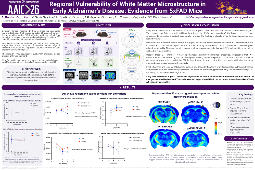
Diffusion Tensor Imaging (DTI) is a magnetic resonance imaging technique that measures water diffusion within brain tissue. In white matter (WM), diffusion patterns reflect fiber organization, axonal structure, and myelin integrity, making DTI useful for detecting microstructural alterations. In Alzheimer’s disease, WM changes may appear during early stages and disrupt structural connectivity between regions involved in memory and cognition, potentially before evident cognitive deficits emerge. Therefore, DTI may help identify subtle WM alterations before overt cognitive impairment.
Aim:
To identify early genotype-, age-, and sex-related changes in WM microstructure in the 5xFAD mouse model using DTI.
Hypothesis:
Diffusion tensor imaging will detect early white matter microstructural alterations in 5xFAD mice before evident cognitive deficits, with differences influenced by age and sex.
Methods:
Male and female WT and 5xFAD mice were assessed longitudinally using diffusion MRI and Y-maze spontaneous alternation. Regions of interest included frontal, medial, and caudal corpus callosum, fimbria, and hippocampus. DTI metrics included FA, RD, AD, and ADC.
Results:
Y-maze performance was preserved across sex, genotype, and age. DTI showed region- and sex-dependent WM alterations. Male 5xFAD mice showed early callosal and fimbrial WM alterations, including reduced FA in the frontal corpus callosum and increased RD in the frontal corpus callosum and fimbria. Female 5xFAD mice showed a distinct age-dependent FA pattern in the medial corpus callosum. Representative FA maps suggested sex-dependent WM organization.
Discussion and Conclusion:
Early WM microstructural alterations were detected in 5xFAD mice, mainly in frontal callosal and fimbrial regions. This regional specificity may reflect differential vulnerability of WM tracts in early Alzheimer’s disease. Reduced FA suggests decreased fiber coherence or altered WM organization, while increased RD may reflect altered radial diffusion and possible myelin-related vulnerability. Despite these DTI changes, Y-maze spontaneous alternation remained preserved, suggesting that WM microstructural alterations may precede overt spatial working memory impairment. FA maps and regional DTI changes suggest sex-dependent patterns of WM organization. Early WM alterations in 5xFAD mice were region-specific and may follow sex-dependent patterns. These DTI changes occurred before overt Y-maze impairment, supporting WM microstructure as a sensitive marker of early AD-related vulnerability.
Key findings:
DTI detected early WM vulnerability in 5xFAD mice.
Frontal CC and fimbria showed selective susceptibility.
Alterations were more evident in male 5xFAD mice.
FA maps suggest sex-dependent WM organization.
References:
1. Maharjan S, et al. Front Neurosci. 2022;16:964654.
2. Molinuevo JL, et al. Neurobiol Aging. 2014;35:2671–2680.
3. Lawrence KE, et al. Brain Imaging Behav. 2021;15:2813–2823.
4. Zhou CN, et al. Front Aging Neurosci. 2018;10:243.
Comments
Email is required but is not shown publicly.
Loading comments...