4,068 posters, 219 videos, 101 topics, 69 sessions, 14,365 authors, 6,128 institutions
ePostersLive by SciGen Technologies S.A. All rights reserved.
Alzheimer's Association International Conference 2026
July 12-15, 2026 | London, United Kingdom, and Online

7316
Regional Specificity Of Enlarged Perivascular Spaces: Temporal Burden Correlates With Amyloid Pathology
Poster Presenter
Authors
Ying Xia, Hang Min, Vincent Doré, Ishara Paranawithana, Yichi Zhang, Fang Ji, Maria del C. Valdés Hernández, Juan Helen Zhou, Jurgen Fripp, Pierrick Bourgeat
Affiliations
The Australian e-Health Research Centre, CSIRO Health and Biosecurity, Brisbane, QLD, Australia, Yong Loo Lin School of Medicine, National University of Singapore, Singapore, Centre for Clinical Brain Sciences, UK Dementia Research Centre, The University of Edinburgh, Edinburgh, UK
Part of Topic
Regional Specificity of Enlarged Perivascular Spaces: Temporal Burden Correlates with Amyloid Pathology
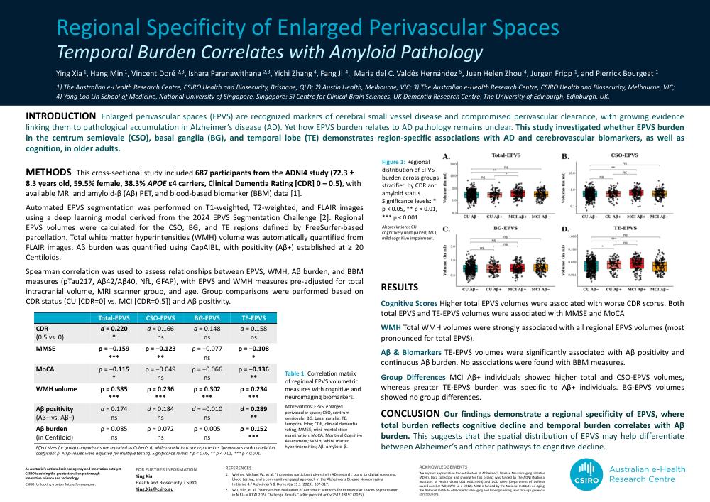
INTRODUCTION Enlarged perivascular spaces (EPVS) are recognized markers of cerebral small vessel disease and compromised perivascular clearance, with growing evidence linking them to pathological accumulation in Alzheimer’s disease (AD). Yet how EPVS burden relates to AD pathology remains unclear. This study investigated whether EPVS burden in the centrum semiovale (CSO), basal ganglia (BG), and temporal lobe (TE) demonstrates region-specific associations with AD and cerebrovascular biomarkers, as well as cognition, in older adults.
METHODS This cross-sectional study included 687 participants from the ADNI4 study (72.3 ± 8.3 years old, 59.5% female, 38.3% APOE ε4 carriers, Clinical Dementia Rating [CDR] 0 – 0.5), with available MRI and amyloid-β (Aβ) PET, and blood-based biomarker (BBM) data [1].
Automated EPVS segmentation was performed on T1-weighted, T2-weighted, and FLAIR images using a deep learning model derived from the 2024 EPVS Segmentation Challenge [2]. Regional EPVS volumes were calculated for the CSO, BG, and TE regions defined by FreeSurfer-based parcellation. Total white matter hyperintensities (WMH) volume was automatically quantified from FLAIR images. Aβ burden was quantified using CapAIBL, with positivity (Aβ+) established at ≥ 20 Centiloids.
Spearman correlation was used to assess relationships between EPVS, WMH, Aβ burden, and BBM measures (pTau217, Aβ42/Aβ40, NfL, GFAP), with EPVS and WMH measures pre-adjusted for total intracranial volume, MRI scanner group, and age. Group comparisons were performed based on CDR status (CU [CDR=0] vs. MCI [CDR=0.5]) and Aβ positivity.
RESULTS
Cognitive Scores Higher total EPVS volumes were associated with worse CDR scores. Both total EPVS and TE-EPVS volumes were associated with MMSE and MoCA.
WMH Total WMH volumes were strongly associated with all regional EPVS volumes (most pronounced for total EPVS).
Aβ & Biomarkers TE-EPVS volumes were significantly associated with Aβ positivity and continuous Aβ burden. No associations were found with BBM measures.
Group Differences MCI Aβ+ individuals showed higher total and CSO-EPVS volumes, whereas greater TE-EPVS burden was specific to Aβ+ individuals. BG-EPVS volumes showed no group differences.
CONCLUSION Our findings demonstrate a regional specificity of EPVS, where total burden reflects cognitive decline and temporal burden correlates with Aβ burden. This suggests that the spatial distribution of EPVS may help differentiate between Alzheimer’s and other pathways to cognitive decline.
Comments
Email is required but is not shown publicly.
Loading comments...