4,068 posters, 219 videos, 101 topics, 69 sessions, 14,365 authors, 6,128 institutions
ePostersLive by SciGen Technologies S.A. All rights reserved.
Alzheimer's Association International Conference 2026
July 12-15, 2026 | London, United Kingdom, and Online

9557
Layer Specific Retinal Thickness As A Marker Of Cerebral Amyloid Burden
Poster Presenter
Part of Topic
Layer Specific Retinal Thickness as a Marker of Cerebral Amyloid Burden
Macular OCT reveals a depth-dependent retinal signature of cerebral amyloid: inner layers thin while outer layers thicken with increasing amyloid burden
Background:
o AD pathology begins decades before symptoms, creating demand for early, scalable, non-invasive biomarkers.
o PET/CSF quantify amyloid but are invasive, costly, and unsuitable for broad screening.
o The retina, as a CNS extension, shows amyloid deposition and structural/vascular changes detectable in vivo.
o OCT provides rapid, reproducible, layer-resolved metrics linked to AD-related neurodegeneration, though findings vary across studies.
o Few studies have related retinal structure to quantitative cerebral amyloid burden (centiloid) in cognitively unimpaired individuals.
Aim:
o To determine whether layer and regionspecific retinal thickness is associated with cerebral amyloid burden in cognitively unimpaired adults.
Methods:
Participants
o 52 cognitively unimpaired adults
o Aβ status: 16 Aβ+, 36 Aβ
Imaging
o PET: [18F-Florbetaben]; global amyloid burden (centiloid)
o OCT: SD-OCT macular & optic-disc scans (OD/OS)
o Retinal metrics: Layer-resolved thickness (ETDRS subfields)
Results:
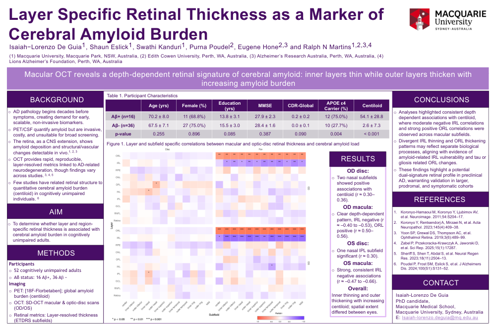
o OD disc: Two nasal subfields showed positive associations with centiloid (r ≈ 0.300.36).
o OD macula: Clear depth-dependent pattern, IRL negative (r ≈ –0.40 to –0.53), ORL positive (r ≈ 0.500.56).
o OS disc: One nasal IPL subfield significant (r ≈ 0.30).
o OS macula: Strong, consistent IRL negative associations (r ≈ –0.47 to –0.66).
o Overall: Inner thinning and outer thickening with increasing centiloid; spatial extent differed between eyes.
Conclusions:
o Analyses highlighted consistent depth dependent associations with centiloid, where moderate negative IRL correlations and strong positive ORL correlations were observed across macular subfields.
o Divergent IRL thinning and ORL thickening patterns may reflect separate biological processes, aligning with evidence of amyloid-related IRL vulnerability and tau or gliosis related ORL changes.
o These findings highlight a potential dual-signature retinal profile in preclinical AD, warranting validation in larger, prodromal, and symptomatic cohorts
Comments
Email is required but is not shown publicly.
Loading comments...