4,068 posters, 219 videos, 101 topics, 69 sessions, 14,365 authors, 6,128 institutions
ePostersLive by SciGen Technologies S.A. All rights reserved.
Alzheimer's Association International Conference 2026
July 12-15, 2026 | London, United Kingdom, and Online

6553
Investigating The Interaction Of Apoe4 With Tdp43 Mislocalisation In Driving Neurodegeneration
Poster Presenter
Authors
Affiliations
Part of Topic
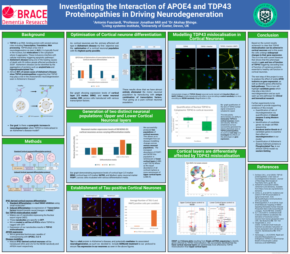
Investigating the interaction of APOE4 and TDP43 proteinopathies in Driving Neurodegeneration Antonio Fusciardi, Professor Jonathan Mill and Dr Akshay Bhinge. Living systems institute, University of Exeter, Devon, UK
Background ● TDP43 is an RNA binding protein with several various roles including Transcription, Translation, RNA processing and many more. TDP43 plays a key role in neurodegeneration as seen in ALS. In ALS the TDP43, which is typically found in the nucleus, mislocalises to the cytoplasm where it aggregates. These inclusions interfere with cellular machinery triggering apoptotic pathways 1 . ● Alzheimer's disease being one of the leading causes of death with 55 million people affected worldwide in 20202 . Alzheimer's disease can be identified by the aggregation of hallmark proteins such as amyloid beta and Hyperphosphorylated Tau. ● Interestingly almost 80% of severe cases of Alzheimer's Disease show TDP43 proteinopathies suggesting that TDP43 may play a role in the characteristic neurodegeneration seen in Alzheimer's Disease 3 . ● Our goal: Is there a synergistic increase in neurodegeneration when TDP43 is mislocalised in an Alzheimer's disease model?
What is the current standard protocol for IPSC-derived Cortical Neurone differentiation? Currently there are two methods by which most groups generate IPSC-derived cortical neurones 1. Standard differentiation via dual SMAD inhibition using small molecules 4 2. Induced differentiation via expression of Transcription factors biased towards neural lineages ie NGN2. 5 We explore the specifics of each protocol in our "methods" section
Methods FNPC generation Cortical neurone differentiation Cortical neurone maturation Disease modelling D0 D18 D25 D38 D58: Collection Cell plating Cortical neurone differentiation Cortical neurone maturation Disease modelling D-2 D0 D12 D23 D43: Collection NGN2 transduction Induced Cortical neuronal differentiation protocol Standard Cortical neuronal differentiation protocol Our TDP43-mislocalisation model 7 : ● Makes use of nanobodies expressing the Nuclear export signal (NES). ● These nanobodies are specific to GFP. ● We've created a cell line of IPSCs where TDP43 is tagged with GFP. ● Expression of our nanobodies in these cells results in TDP43 mislocalisation to the cytoplasm. APOE4 expression ● We've generated several lentiviruses capable of transducing cells with either APOE2, 3 or 4. Disease modelling ● Mature IPSC-derived cortical neurones will be transduced AAVs and LVs for the NESM nanobody and APOE4 insert respectively.
Establishment of Tau-positive cortical neurones: Tau is a vital protein in Alzheimer's disease, and potentially mediates its associated neurodegeneration, as such we decided to include GENtonik treatment to our protocol to ensure Tau expression in our neurones as seen in the adjacent figure.
Optimisation of Cortical neurone differentiation: Bar graph demonstrating various expression levels of cortical layer 2/3 marker, BRN2, and motor neuronal marker, HB9, across cells transduced with various transcription factors.
Generation of two distinct neuronal populations: Upper and Lower cortical layers Bar graph demonstrating various expression levels of cortical layer 2/3 marker BRN2, cortical layer 4/5 marker SATB2, and Medium spiny neuronal marker FOXP1, across cells incubated with various differentiation media. We successfully produced two populations of cortical neurones. This gives us the opportunity to look at how our disease modelling changes across various neuronal populations. Due to the increased abundance of FOXP1 and SATB2 this population is reminiscent of lower cortical layers (LSBE-x-pd173074) Where as the equal abundance of all cortical markers is more reminiscent of Upper cortical layers (LSBE-X).
Modelling TDP43 mislocalisation in cortical neurones
immunostain images of TDP43 (Green) neuronal nuclei stained with Hoechst (Blue), and MAP2 (Yellow) in cells transduced with either a virus encoding a control nanobody or a virus encoding our NESM nanobody. Magnification is 20x.
Cortical layers are differentially affected by TDP43 mislocalisation: UMAP and Volcano plots resulting from Single cell RNA sequencing to identify candidate genes affected by TDP43 mislocalisation. We successfully identified that the lower cortical layers of the cortex are substantially more affected by TDP43 mislocalisation than Upper cortical layers.
Future work In the future to answer many of our remaining questions we would need further work to: ● Quantification of rate of neurodegeneration via quantification of cleaved caspase 3 using Western Blotting ● Identification of candidate genes via single cell RNA sequencing. ● Knockout and/or Knock-in of candidate genes to examine effect on neurodegeneration in our model. ● Quantifcation of Alzheimer's disease hallmark proteins ie Phosphorylated Tau, in our APOE4-TDP43 model via western blotting.
Comments
Email is required but is not shown publicly.
Loading comments...