4,068 posters, 219 videos, 101 topics, 69 sessions, 14,365 authors, 6,128 institutions
ePostersLive by SciGen Technologies S.A. All rights reserved.
Alzheimer's Association International Conference 2026
July 12-15, 2026 | London, United Kingdom, and Online

9645
Clinicopathological Correlates Of FTLD-TDP Type E: A Deep Phenotyping And Autopsy Case Report
Poster Presenter
Authors
Sara Rubio-Guerra, Jesús García Castro, Judit Selma-González, Isabel Sala, Mª Belén Sánchez-Saudinós, Nuole Zhu, Isabel Barroeta, Ricardo Rojas-Garcia, Janina Turon-Sans, Álvaro Carbayo, Valle Camacho, David Almenta, Josefina Pérez-Blanco, Sara Bernal, Lidia González-Quereda, Laura Molina-Porcel, Oriol Dols-Icardo, Daniel Alcolea, Juan Fortea, Alberto Lleó, Iban Aldecoa, Ignacio Illán-Gala
Affiliations
Sant Pau Memory Unit, Department of Neurology, IR SANT PAU, Hospital de la Santa Creu i Sant Pau, Barcelona, Spain; Center for Biomedical Network Research on Neurodegenerative Diseases (CIBERNED), Instituto de Salud Carlos III, Madrid, Spain; Department of Medicine, Universitat Autònoma de Barcelona, Barcelona, Spain, Sant Pau Memory Unit, Department of Neurology, IR SANT PAU, Hospital de la Santa Creu i Sant Pau, Barcelona, Spain; Center for Biomedical Network Research on Neurodegenerative Diseases (CIBERNED), Instituto de Salud Carlos III, Madrid, Spain; Institute of Neurosciences, Universitat Autònoma de Barcelona, Barcelona, Spain, Sant Pau Memory Unit, Department of Neurology, IR SANT PAU, Hospital de la Santa Creu i Sant Pau, Barcelona, Spain; Center for Biomedical Network Research on Neurodegenerative Diseases (CIBERNED), Instituto de Salud Carlos III, Madrid, Spain, Motor Neuron Disease Clinic, Neuromuscular Diseases Unit, Department of Neurology, IR SANT PAU, Hospital de la Santa Creu i Sant Pau, Barcelona, Spain, Department of Nuclear Medicine, IR SANT PAU, Hospital de la Santa Creu i Sant Pau, Barcelona, Spain, Department of Psychiatry, IR SANT PAU, Hospital de la Santa Creu i Sant Pau, Barcelona, Spain, Department of Genetics, IR SANT PAU, Hospital de la Santa Creu i Sant Pau, Barcelona, Spain; Center for Biomedical Network Research on Rare Diseases (CIBERER), Instituto de Salud Carlos III, Madrid, Spain, Neurological Tissue Bank, Biobanc-Hospital Clínic, Hospital Clínic de Barcelona, Fundació de Recerca Clínic Barcelona (FRCB)–Institut d’Investigacions Biomèdiques August Pi I Sunyer (IDIBAPS), Barcelona, Spain; Alzheimer’s disease and other cognitive disorders Unit, Hospital Clínic de Barcelona, Fundació de Recerca Clínic Barcelona (FRCB)–Institut d’Investigacions Biomèdiques August Pi i Sunyer (IDIBAPS), Barcelona, Spain, Neurological Tissue Bank, Biobanc-Hospital Clínic, Hospital Clínic de Barcelona, Fundació de Recerca Clínic Barcelona (FRCB)–Institut d’Investigacions Biomèdiques August Pi I Sunyer (IDIBAPS), Barcelona, Spain; Department of Pathology, Biomedical Diagnostic Center, Hospital Clínic de Barcelona, Barcelona, Spain
Part of Topic
BACKGROUND
Nearly half of patients with frontotemporal lobar degeneration (FTLD) have underlying TDP-43 pathology (FTLD-TDP). FTLD-TDP is subclassified into types A to D according to the morphology and distribution of TDP-43 aggregates.(1) Recently, FTLD-TDP pathological classification was updated to include a fifth subtype (TDP-43 type E) associated with motor neuron involvement and rapid clinical progression.(2) However, these clinical features and similarities between FTLD-TDP type E pathological findings and those of FTLD-TDP type B have raised the question of whether FTLD-TDP type E might actually represent an early form of or be related to FTLD-TDP type B.(3) To date, few cases of FTLD-TDP type E have been reported.(2,4,5)
METHODS
We describe the clinical and neuropathological findings in a patient with FTLD-TDP type E. The clinical examination comprised electromyography, [¹⁸F]fluorodeoxyglucose positron emission tomography-computed tomography ([¹⁸F]FDG-PET/CT), laboratory workup, cerebrospinal fluid (CSF) Alzheimer’s disease (AD) biomarker analysis, testing for C9orf72, and an FTLD-targeted gene panel (n=108). A neuropsychological evaluation and brain magnetic resonance imaging (MRI) had previously been conducted at another center.
RESULTS
A right-handed 68-year-old woman with secondary education presented with a 4-year history of insidiously progressive personality change: the initial symptom, as reported by her husband, was behavioral disinhibition (including impulsive actions, socially inappropriate behavior, and loss of manners), soon followed by apathy and inertia, loss of empathy, motor and speech stereotypies, binge eating, and compulsive smoking and consumption of alcohol and sunflower seeds. The informant also reported memory deficits, word-finding difficulties, formed visual hallucinations, pareidolias, and false recognition of strangers as familiar people. Over the course of the disease, the patient also developed progressive motor impairment with marked gait slowing, emotional lability, dysphagia, and urge urinary incontinence. The patient was unaware of most of these changes. At initial assessment, she was dependent for basic activities of daily living. Family history was negative for neurodegenerative or psychiatric disorders.
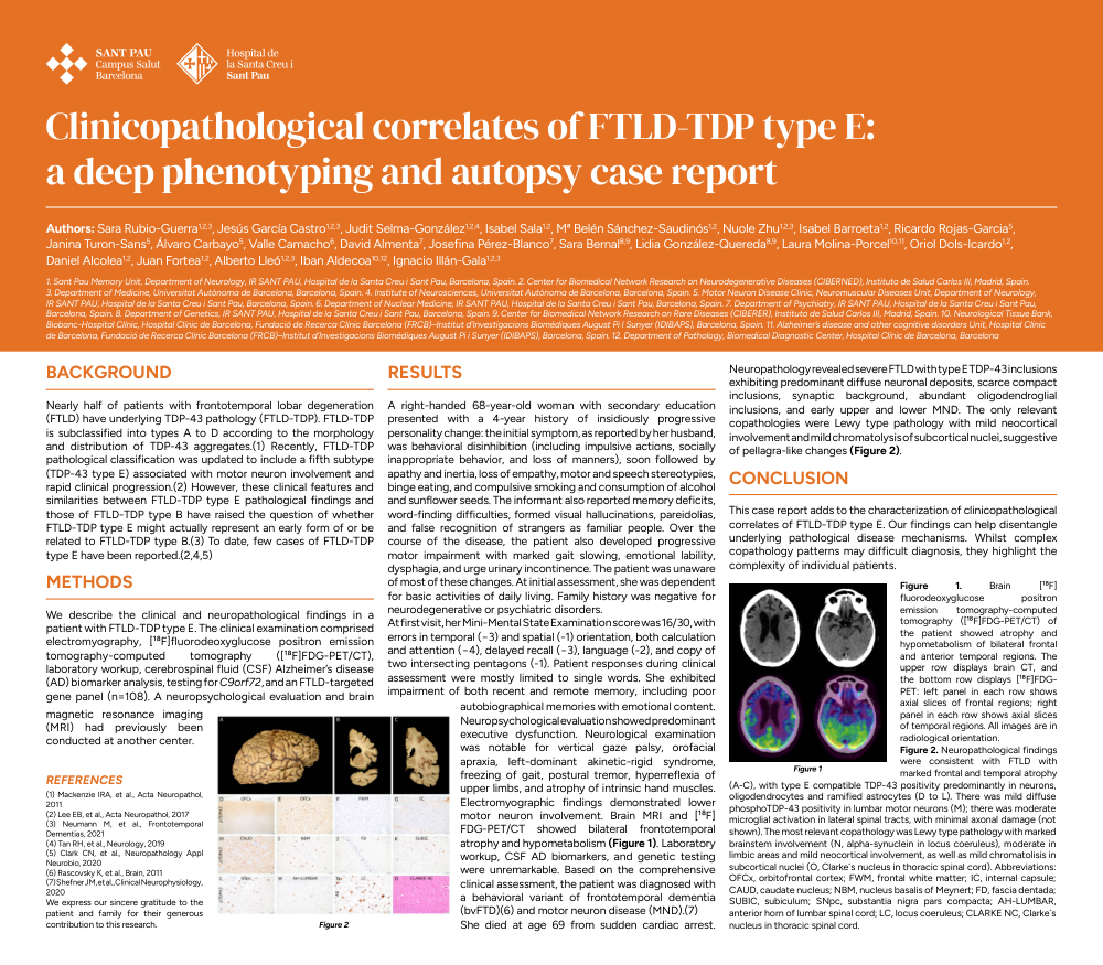
At first visit, her Mini-Mental State Examination score was 16/30, with errors in temporal (−3) and spatial (-1) orientation, both calculation and attention (−4), delayed recall (−3), language (-2), and copy of two intersecting pentagons (-1). Patient responses during clinical assessment were mostly limited to single words. She exhibited impairment of both recent and remote memory, including poor autobiographical memories with emotional content. Neuropsychological evaluation showed predominant executive dysfunction. Neurological examination was notable for vertical gaze palsy, orofacial apraxia, left-dominant akinetic-rigid syndrome, freezing of gait, postural tremor, hyperreflexia of upper limbs, and atrophy of intrinsic hand muscles. Electromyographic findings demonstrated lower motor neuron involvement. Brain MRI and [¹⁸F]FDG-PET/CT showed bilateral frontotemporal atrophy and hypometabolism (Figure 1). Laboratory workup, CSF AD biomarkers, and genetic testing were unremarkable. Based on the comprehensive clinical assessment, the patient was diagnosed with a behavioral variant of frontotemporal dementia (bvFTD)(6) and motor neuron disease (MND).(7)
She died at age 69 from sudden cardiac arrest. Neuropathology revealed severe FTLD with type E TDP-43 inclusions exhibiting predominant diffuse neuronal deposits, scarce compact inclusions, synaptic background, abundant oligodendroglial inclusions, and early upper and lower MND. The only relevant copathologies were Lewy type pathology with mild neocortical involvement and mild chromatolysis of subcortical nuclei, suggestive of pellagra-like changes (Figure 2).
CONCLUSION
This case report adds to the characterization of clinicopathological correlates of FTLD-TDP type E. Our findings can help disentangle underlying pathological disease mechanisms. Whilst complex copathology patterns may difficult diagnosis, they highlight the complexity of individual patients.
Figure 1. Brain [¹⁸F]fluorodeoxyglucose positron emission tomography-computed tomography ([¹⁸F]FDG-PET/CT) of the patient showed atrophy and hypometabolism of bilateral frontal and anterior temporal regions. The upper row displays brain CT, and the bottom row displays [¹⁸F]FDG-PET: left panel in each row shows axial slices of frontal regions; right panel in each row shows axial slices of temporal regions. All images are in radiological orientation.
Figure 2. Neuropathological findings were consistent with FTLD with marked frontal and temporal atrophy (A-C), with type E compatible TDP-43 positivity predominantly in neurons, oligodendrocytes and ramified astrocytes (D to L). There was mild diffuse phosphoTDP-43 positivity in lumbar motor neurons (M); there was moderate microglial activation in lateral spinal tracts, with minimal axonal damage (not shown). The most relevant copathology was Lewy type pathology with marked brainstem involvement (N, alpha-synuclein in locus coeruleus), moderate in limbic areas and mild neocortical involvement, as well as mild chromatolisis in subcortical nuclei (O, Clarke´s nucleus in thoracic spinal cord). Abbreviations: OFCx, orbitofrontal cortex; FWM, frontal white matter; IC, internal capsule; CAUD, caudate nucleus; NBM, nucleus basalis of Meynert; FD, fascia dentada; SUBIC, subiculum; SNpc, substantia nigra pars compacta; AH-LUMBAR, anterior horn of lumbar spinal cord; LC, locus coeruleus; CLARKE NC, Clarke´s nucleus in thoracic spinal cord.
REFERENCES
(1) Mackenzie IRA, et al., Acta Neuropathol, 2011
(2) Lee EB, et al., Acta Neuropathol, 2017
(3) Neumann M, et al., Frontotemporal Dementias, 2021
(4) Tan RH, et al., Neurology, 2019
(5) Clark CN, et al., Neuropathology Appl Neurobio, 2020
(6) Rascovsky K, et al., Brain, 2011
(7) Shefner JM, et al., Clinical Neurophysiology, 2020
We express our sincere gratitude to the patient and family for their generous contribution to this research.
Comments
Email is required but is not shown publicly.
Loading comments...