4,068 posters, 219 videos, 101 topics, 69 sessions, 14,365 authors, 6,128 institutions
ePostersLive by SciGen Technologies S.A. All rights reserved.
Alzheimer's Association International Conference 2026
July 12-15, 2026 | London, United Kingdom, and Online

1616
Left-Predominant Hippocampal Atrophy Associated With LATE-NC
Poster Presenter
Authors
Lama Rayyis, Hyunwoo Lee, Waad Alshammari, Julia Xie, Ian R Mackenzie, Ging-Yuek Robin Hsiung
Affiliations
University of British Columbia, Vancouver, BC, Canada, Division of Neurology, University of British Columbia, Vancouver, BC, Canada, 2University of British Columbia, Vancouver, BC, Canada, Department of Pathology & Laboratory Medicine, University of British Columbia, Vancouver, BC, Canada.
Part of Topic
Left-Predominant Hippocampal Atrophy Associated With LATE-NC
Lama Rayyis1, Hyunwoo Lee1, Waad Alshammari1, Julia Xie2, Ian R. Mackenzie3
Ging-Yuek Robin Hsiung1
1 Division of Neurology, The University of British Columbia, Vancouver, BC, Canada
2 The University of British Columbia, Vancouver, BC, Canada
3 Department of Pathology & Laboratory Medicine, University of British Columbia, Vancouver, BC, Canada
Background:
Limbic-predominant age-related TDP-43 encephalopathy (LATE) causes an amnestic syndrome that closely mimics Alzheimer’s disease and commonly co-occurs with it. No validated in vivo biomarkers exist for TDP-43 pathology yet, and diagnosis is confirmed postmortem. MRI-based surrogate markers may help identify limbic TDP-43 involvement during life. Prior autopsy and MRI studies suggest that a higher middle–inferior temporal gyrus (MITG)–to–hippocampus volume ratio may distinguish TDP-43 pathology from AD. We evaluated MRI volumetric measures in our autopsy-confirmed cohort with mixed dementias and LATE neuropathologic change (LATE-NC).
Methods:
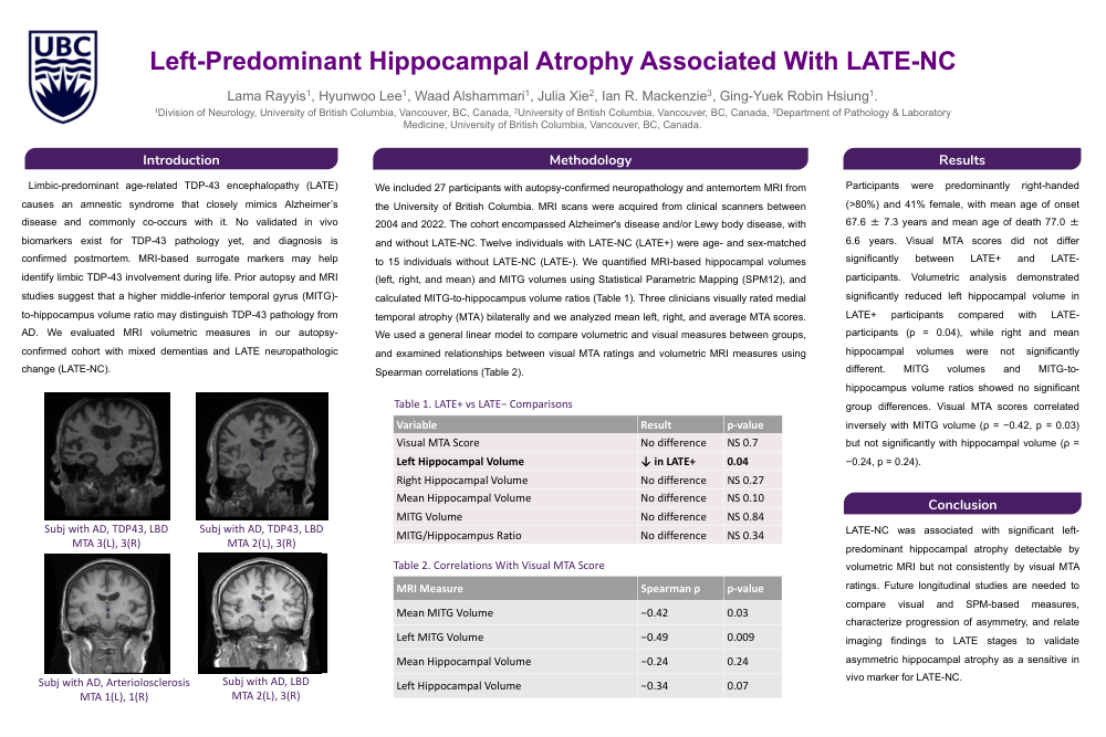
We included 27 participants with autopsy-confirmed neuropathology and antemortem MRI from the University of British Columbia. MRI scans were acquired from clinical scanners between 2004 and 2022. The cohort encompassed Alzheimer's disease and/or Lewy body disease, with and without LATE-NC. Twelve individuals with LATE-NC (LATE+) were age- and sex-matched to 15 individuals without LATE-NC (LATE-). We quantified MRI-based hippocampal volumes (left, right, and mean) and MITG volumes using Statistical Parametric Mapping (SPM12), and calculated MITG-to-hippocampus volume ratios (Table 1). Three clinicians visually rated medial temporal atrophy (MTA) bilaterally and we analyzed mean left, right, and average MTA scores. We used a general linear model to compare volumetric and visual measures between groups, and examined relationships between visual MTA ratings and volumetric MRI measures using Spearman correlations (Table 2).
Results:
Participants were predominantly right-handed (>80%) and 41% female, with mean age of onset 67.6 ± 7.3 years and mean age of death 77.0 ± 6.6 years. Visual MTA scores did not differ significantly between LATE+ and LATE- participants. Volumetric analysis demonstrated significantly reduced left hippocampal volume in LATE+ participants compared with LATE- participants (p = 0.04), while right and mean hippocampal volumes were not significantly different. MITG volumes and MITG-to- hippocampus volume ratios showed no significant group differences. Visual MTA scores correlated inversely with MITG volume (ρ = −0.42, p = 0.03) but not significantly with hippocampal volume (ρ = −0.24, p = 0.24).
Conclusion:
LATE-NC was associated with significant left-predominant hippocampal atrophy detectable by volumetric MRI but not consistently by visual MTA ratings. Future longitudinal studies are needed to compare visual and SPM-based measures, characterize progression of asymmetry, and relate imaging findings to LATE stages to validate asymmetric hippocampal atrophy as a sensitive in vivo marker for LATE-NC.
Comments
Email is required but is not shown publicly.
Loading comments...