4,068 posters, 219 videos, 101 topics, 69 sessions, 14,365 authors, 6,128 institutions
ePostersLive by SciGen Technologies S.A. All rights reserved.
Alzheimer's Association International Conference 2026
July 12-15, 2026 | London, United Kingdom, and Online

467
Immune–epithelial remodeling axis in the human choroid plexus in Alzheimer’s disease
Poster Presenter
Authors
Jaroslav Bendl, Milos Pjanic, Seon Kinrot, Xinyi Wang, Xuan Cao, Christian Porras, Prashant N M, Sarah Murphy, Annie Khamhoung, Tereza Clarence, Clara Casey, Aram Hong, Zhiping Shao, Chenyu Liu, Deepika Mathur, Maxim Signaevski, Georgios Voloudakis, Gabriel Hoffman, Cameron McAlpine, Vahram Haroutunian, Donghoon Lee, John Fullard, Jaroslav Bendl, Panos Roussos
Affiliations
Part of Topic
Immune–epithelial remodeling axis in the human choroid plexus in Alzheimer’s disease
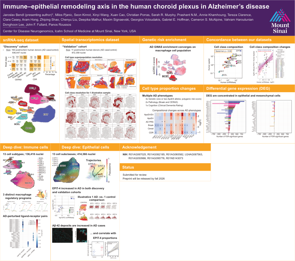
Background: The choroid plexus (CP) regulates cerebrospinal fluid composition and immune surveillance at the brain’s borders, but its contribution to AD is not well characterized. In particular, it remains unknown how alterations within the human CP relate to AD severity and downstream pathological features.
Methods: We profiled 936,447 nuclei from 160 postmortem human CP samples using single-nucleus RNA sequencing (snRNA-seq), spanning neurotypical controls and AD, to construct a high-resolution cellular atlas (16 major classes, 37 subclasses, 64 subtypes). We integrated genetic risk mapping (heritability enrichment), compositional modeling across AD genetic, clinical, and neuropathological measures, differential expression and regulatory network analyses, cell–cell interaction inference, and causal mediation modeling. Spatial transcriptomics at subcellular resolution (4 early AD, 4 late AD, 4 controls; 5,001-gene panel) provided in situ validation.
Results: AD heritability enrichment concentrated in CP immune populations, most prominently macrophages. Across genetic, clinical, and neuropathological measures of AD, we observed progressive depletion of regulatory immune states (including anti-inflammatory CD8⁺ T cells) alongside accumulation of pro-inflammatory macrophages. Spatial profiling revealed macrophage expansion in the parenchyma (≈5% in controls to ≈15% in late AD) and an increased pro-/anti-inflammatory macrophage ratio in AD, detected in both the CP stroma and parenchyma.This inflammatory skew associated with closer macrophage proximity to epithelial cells, consistent with heightened immune pressure at the barrier interface.
Epithelial states followed disease-linked transitions: as AD advances, late-stage epithelium exhibited marked loss of transthyretin (TTR), activation of stress and DNA-damage programs, and structural remodeling consistent with epithelial thickening, alongside a distinct epithelial-to-mesenchymal transition (EMT)–like program with ZEB1, TGF-β, and WNT signaling as candidate drivers. These diseased epithelial signatures were supported by spatial transcriptomics and replicated in independent CP datasets and in a PS19 tauopathy mouse model.
Mediation modeling supported an immune-driven disease cascade in which AD-associated immune perturbations are linked to epithelial injury, which in turn precedes and partially explains downstream amyloid and tau pathology.
Conclusion: Our results position the CP as an active immune–barrier nexus in AD, where macrophage-centered immune remodeling is coupled to epithelial dysfunction and EMT-like reprogramming.
Comments
Email is required but is not shown publicly.
Loading comments...