4,068 posters, 219 videos, 101 topics, 69 sessions, 14,365 authors, 6,128 institutions
ePostersLive by SciGen Technologies S.A. All rights reserved.
Alzheimer's Association International Conference 2026
July 12-15, 2026 | London, United Kingdom, and Online

4848
Association Between Amyloid Pet And Hippocampus Atrophy In A Real-world Cohort: Preliminary Findings
Poster Presenter
Authors
Bruna Bressan Valentini, Anelise Vivian Fuhr, Brunna Teló Jaeger, André Luiz Rodrigues Palmeira, Renata Kochhann, Joana Emilia Senger, Mariana Dagnino, Sheila Cristina Ouriques Martins, Luiz Antônio Nasi, Eduardo Rigon Zimmer, Pedro Rosa-Neto, Wyllians Vendramini Borelli
Affiliations
Universidade Federal do Rio Grande do Sul; Hospital Moinhos de Vento, Hospital Moinhos de Vento, Universidade Federal do Rio Grande do Sul; Hospital Moinhos de Vento; McGill University, McGill University; University of Texas
Part of Topic
Title: Association between amyloid PET and hippocampus atrophy in a real-world cohort: preliminary findings
Authors:
Bruna Bressan Valentini, MSc student (1, 2); Anelise Vivian Fuhr (2); Brunna Teló Jaeger (2); André Luiz Rodrigues Palmeira (2); Renata Kochhann (2); Joana Senger (1, 2); Mariana Dagnino (2); Sheila Cristina Ouriques Martins (1, 2); Luiz Antônio Nasi (2); Eduardo R. Zimmer (1, 2, 3); Pedro Rosa-Neto (3,4); Wyllians Jose Vendramini Borelli, MD, PhD (1, 2)
Affiliations: (1) Universidade Federal do Rio Grande do Sul, Porto Alegre, Rio Grande do Sul, Brazil, (2) Hospital Moinhos de Vento, Porto Alegre, Rio Grande do Sul, Brazil, (3) McGill University, Montreal, Canada; (4) University of Texas, USA.
Introduction: Current Alzheimer’s disease (AD) models are largely built on homogenous, highly educated, Caucasian cohorts. AD pathology in vivo should also be replicated in resource-limited countries with heterogeneous genetic backgrounds to ensure the validity of the ATN framework globally. This study aimed to test the association between amyloid burden (A) and hippocampal volume (N) in a real-world cohort.
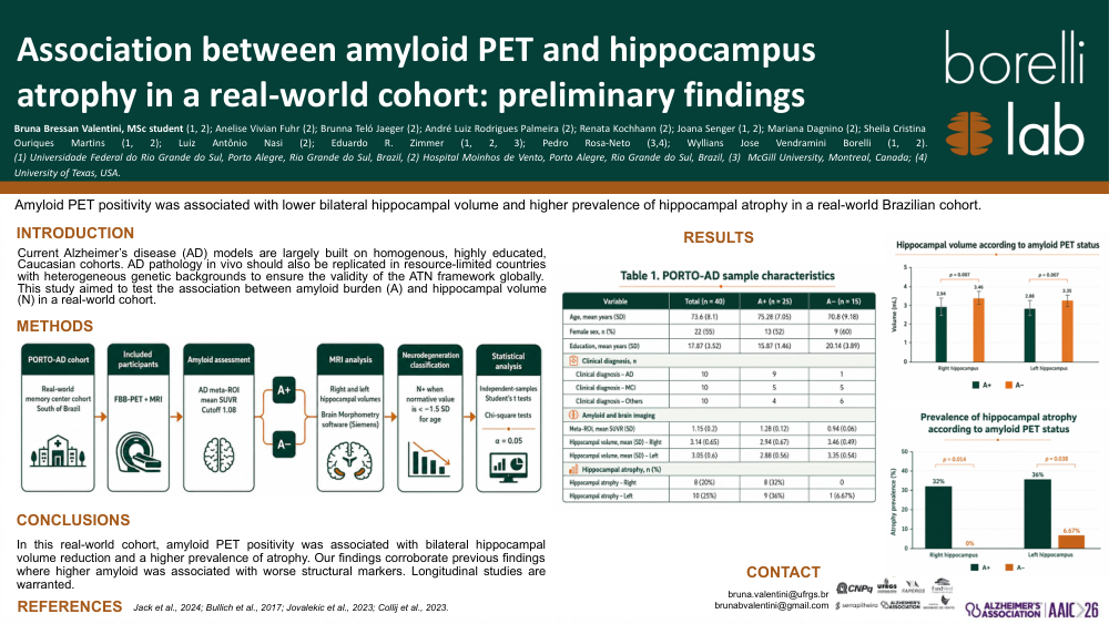
Methods: This is a preliminary analysis of the PORTO-AD cohort - a real-world cohort from a memory center in south of Brazil. Individuals who underwent FBB-PET and MRI were included. The AD meta-ROI mean SUVR extracted from PET was assessed as negative (A-) or positive (A+), using 1.08 as the cutoff. Right and left hippocampal volumes (mL) were compared using Brain Morphometry software (Siemens) and individuals were classified as N+ when normative values were -1.5 SD than mean for age. Hippocampal atrophy was compared across A+ and A- groups. Independent-samples Student’s t tests and chi-square association tests were applied (α=0.05) accordingly.
Results: Forty cases were analyzed (Table 1). The A+ group showed lower hippocampal volume. Right hippocampal volume was 3.46 mL (0.494) in the A- group versus 2.94 mL (0.666) in the A+ group (t(38)=2.60; p=0.007). Left hippocampal volume was 3.35 mL (0.544) in A- versus 2.88 mL (0.563) in A+ (t(38)=2.59; p=0.007). The proportion of N+ was higher in the A+ group when compared with the A- group for right (χ²=6.00; p=0.014) and left hippocampus (χ²=4.30; p=0.038).
Conclusion: In this real-world cohort, amyloid PET positivity was associated with bilateral hippocampal volume reduction and a higher prevalence of atrophy. Our findings corroborate previous findings where higher amyloid was associated with worse structural markers. Longitudinal studies are warranted.
Comments
Email is required but is not shown publicly.
Loading comments...