4,068 posters, 219 videos, 101 topics, 69 sessions, 14,365 authors, 6,128 institutions
ePostersLive by SciGen Technologies S.A. All rights reserved.
Alzheimer's Association International Conference 2026
July 12-15, 2026 | London, United Kingdom, and Online

10388
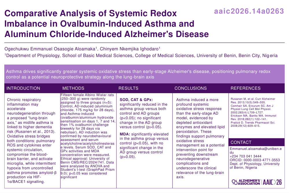
Comparative Analysis Of Systemic Redox Imbalance In Ovalbumin-induced Asthma And Aluminum Chloride-induced Alzheimer's Disease.
Poster Presenter
Authors
Affiliations
Part of Topic
Comparative Analysis of Systemic Redox Imbalance in Ovalbumin-Induced Asthma and Aluminum Chloride-Induced Alzheimer’s Disease
Ogechukwu Emmanuel Osasogie ALOAMAKA1, Chinyem Nkemjika IGHODARO1,
1Department of Physiology, School of Basic Medical Sciences, College of Medical Sciences, University of Benin. Benin City. Nigeria
Author for correspondence: Aloamaka Ogechukwu Emmanuel Osasogie, Department of Physiology, School of Basic Medical Sciences, University of Benin. Benin City. Nigeria.
Emmanuel.aloamaka@uniben.edu, +2347030413444.
Orcid ID: https://orcid.org/0000-0003-4771-3553
Abstract
Introduction: Emerging evidence suggests a "lung-brain axis" where chronic respiratory inflammation may accelerate neurodegeneration. This study investigated the oxidative stress profiles of bronchial asthma and Alzheimer’s disease (AD) to understand their systemic pathological impact.
Methods: Fifteen female Albino Wistar rats were divided into three groups: Control, AD-induced (175 mg/kg Aluminum chloride for 28 days), and Asthma-induced (1 mg Ovalbumin (OVA) and 20 mg aluminium hydroxide sensitization on days 1, 7, and 14 and 1 mg OVA w/v challenge biweekly for 28 days). Oxidative stress was quantified by measuring the serum activities of Superoxide Dismutase (SOD), Catalase (CAT), and Glutathione Peroxidase (GPx), as well as the concentration of Malondialdehyde (MDA). Data were analyzed using ANOVA and Student’s t-tests.
Results: The asthma-induced group exhibited a statistically significant decrease in SOD, CAT, and GPx activities compared to both the control and AD groups (p<0.05). Additionally, MDA levels, a marker of lipid peroxidation, were significantly elevated in the asthma group. Conversely, the AD-induced group showed no statistically significant alterations in these peripheral oxidative markers compared to controls (p>0.05).
Conclusion: Asthma induces a more profound systemic oxidative stress response than the early-stage AD model used in this study. The significant depletion of antioxidant enzymes and increased lipid peroxidation in asthmatic rats underscore the systemic nature of respiratory inflammation. These findings suggest that managing pulmonary oxidative stress may be a critical intervention point for preventing downstream systemic complications, including neurodegenerative decline.
Keywords: Asthma, Alzheimer’s Disease, Oxidative Stress, Lung-Brain Axis, Neuroinflammation.
INTRODUCTION
Historically, the fields of pulmonology and neurology have operated in relative isolation, treating bronchial asthma and Alzheimer’s disease (AD) as distinct pathologies confined to the respiratory tract and the central nervous system (CNS), respectively. However, this compartmentalized view of human physiology is increasingly being challenged by epidemiological data suggesting a robust comorbidity between chronic respiratory inflammation and neurodegenerative decline. A pivotal longitudinal study by (Rusanen et al., 2013) provided one of the first major signals of this connection, demonstrating that individuals who have midlife asthma are at a significantly higher risk of developing dementia later in life. This correlation implies that the consequences of asthmatic pathology extend far beyond the alveolar level, hinting at a systemic "lung-brain axis" where chronic peripheral distress accelerates central aging. The physiological bridge connecting these disparate conditions appears to rest on the foundation of oxidative stress, a pervasive mechanism of cellular damage that serves as a common denominator in both airway remodeling and neuronal loss.
To understand this connection, one must first consider oxidative stress as a fundamental driver of pathology in both systems. Oxidative stress is defined by an imbalance between the production of reactive oxygen species (ROS) and the body’s ability to detoxify these reactive intermediates through antioxidant defenses. In asthma, the airway epithelium is constantly exposed to a barrage of ROS generated by infiltrating leukocytes, particularly eosinophils and neutrophils, recruited to combat allergens or environmental pollutants (Comhair & Erzurum, 2002). While this "oxidative burst" is intended to neutralize pathogens, its chronic activation results in significant collateral damage to lung tissue. Crucially, this inflammatory storm is not confined to the thoracic cavity; the byproducts of pulmonary oxidation—including pro-inflammatory cytokines such as tumor necrosis factor-alpha (TNF-alpha) and interleukin-1 beta (IL-1\beta)—spill over into the systemic circulation, transforming a local respiratory event into a body-wide inflammatory state.
Once these inflammatory mediators enter the bloodstream, they pose a direct threat to the brain’s protective fortifications. The blood-brain barrier (BBB), traditionally viewed as an impermeable shield, can be compromised by chronic systemic inflammation. Research indicates that circulating cytokines can alter endothelial permeability or undergo transport across the BBB, thereby signaling microglia—the brain’s resident immune cells—to enter a reactive state (Erickson & Banks, 2018). This process, known as neuroinflammation, creates a fertile ground for oxidative stress within the CNS. The brain is uniquely vulnerable to this type of damage due to its disproportionately high oxygen consumption and its abundance of polyunsaturated fatty acids, which are highly susceptible to lipid peroxidation. In the pathogenesis of Alzheimer’s disease, oxidative stress is not merely a secondary symptom but a primary instigator. It has been shown to precede the formation of senile plaques and neurofibrillary tangles, suggesting that ROS-mediated damage promotes the cleavage of the amyloid precursor protein (APP) into toxic amyloid-beta peptides (Praticò, 2008).
Furthermore, the link between asthma and Alzheimer’s is compounded by the mechanical reality of respiratory disease: hypoxia. Severe or uncontrolled asthma leads to intermittent periods of reduced oxygen saturation, creating a state of chronic intermittent hypoxia that mirrors the effects of sleep apnea. The brain, being an obligate aerobe with high metabolic demands, is exquisitely sensitive to fluctuations in oxygen supply. Hypoxia stabilizes Hypoxia-Inducible Factor 1-alpha (HIF-1-alpha), a transcription factor that regulates cellular adaptation to low oxygen. At the same time, adaptive in the short term, chronic activation of HIF-1-alpha has been linked to the upregulation of beta-secretase (BACE1), the rate-limiting enzyme responsible for the production of amyloid-beta (Peers et al., 2007). Consequently, the "air hunger" experienced by asthmatic patients may directly translate into metabolic stress for neurons, thereby accelerating the accumulation of AD-associated biomarkers.
The convergence of these mechanisms suggests a synergistic toxicity where the lungs and brain interact in a deleterious feedback loop. The systemic inflammation and oxidative stress originating in the asthmatic lung weaken the brain's defenses, while intermittent hypoxia lowers the threshold for neuronal injury. This "double hit" hypothesis posits that asthma acts as a peripheral accelerator for central neurodegeneration, particularly in aging populations where antioxidant reserves are already diminishing (Chen et al., 2015). Recognizing this link is clinically vital, as it reframes asthma management not just as a means of improving respiratory comfort, but as a potential preventative strategy for preserving cognitive health. By rigorously controlling pulmonary inflammation and oxidative stress, clinicians may inadvertently be dampening the fires of neurodegeneration, highlighting the critical importance of a holistic approach to chronic disease management.
METHODOLOGY
Experimental Animals
This study used female Albino Wistar rats. The animals received proper care in accordance with international guidelines for the handling of experimental animals. Ethical approval was obtained from the University of Benin, College of Medical Sciences ethics board (CMS/REC/2024/741). The Albino Wistar rats were housed in a clean, calm, and sterile environment at room temperature. They were in cages and had access to food and water ad libitum throughout the experimental process.
Study Design
A total of 15 Albino Wistar rats (250-300 grams) were used for this study. The rats were randomly assigned to 3 groups, with 5 rats per group. Group 1 served as control (water and feed ad libitum), Group 2 (AlCl3 only 100 mg/kg body weight for 28 days), Group 3 (asthma-induced sensitization with 1 mg ovalbumin (OVA) and 20 mg aluminium hydroxide, on day 1, 7, and 14; then challenged with 1% w/v OVA biweekly for 28 days)
Experimental protocol/design
The experiment was carried out in phases
Phase 1
Induction and confirmation of Alzheimer’s disease and asthma
Induction was performed using Aluminium chloride at 175 mg/dl for 28 days (Chen et al., 2021), whilst confirmation of Alzheimer’s induction was conducted in accordance with the methodology employed by Walton (2009). AD was confirmed in groups 2 and 3 using neurobehavioral assessment standards (Enogieru & Omoruyi, 2022), serum acetylcholine and acetylcholinesterase levels (Ellman et al., 1961), and histology (Thakur et al., 2019).
Asthma induction
Asthma was induced according to the guidelines outlined by Thakur et al. (2019). Group 3 was sensitized with 1 mg OVA and 20 mg aluminium hydroxide dissolved in saline on days 1, 7, and 14, and challenged with 1% OVA w/v three times every week for 4 weeks (Thakur et al., 2019). The challenged rats were placed in a plastic chamber measuring 70 cm in diameter and 40 cm in length, connected to a Medel family nebulizer (REF 90543 MEDEL FAMILY SILVER AEROSOL) with aerosol delivery of 0.28 ml/min for 15 min per session.
Test Groups
The rats were randomly assigned to 4 groups, with 10 rats per group.
Group 1: Control
Group 2: AlCl3 only (175mg/kg b.w.)
Group 3: Asthma-induced
Blood sampling and serum isolation
Blood samples were immediately collected from euthanized rats via cardiac puncture and from the portal vein. They were kept at room temperature for 30 min, followed by centrifugation at 5000 rpm (revolutions per minute) for 15 min, and serum was isolated by aspiration. The separated serum was frozen for the subsequent quantitative determination of some biomarkers (Thakur et al., 2019).
Determination of Antioxidant Activity
The catalase activity was measured as described by Cohen et al. (1970). The SOD activity was determined according to the method of Misra and Fridovich (1972). Malondialdehyde concentration was measured as described by Buege and Aust (1978).
Standard statistical software, GraphPad Prism 9.0, was used to analyze the generated data. Results were presented as MEAN ± SEM in tables and/or figures. Bar charts were plotted to show the results. Student's t-test and ANOVA were used to compare results within groups and between groups. P-values less than 0.05 (p<0.05) were taken to be statistically significant.
RESULTS
Alterations in superoxide dismutase in Wistar rats induced with asthma and Alzheimer's disease
The results show a statistically significant decrease in superoxide dismutase enzyme activity in the asthma-induced group compared to control (p<0.05), but no statistically significant difference in the Alzheimer's-induced group compared to control (p>0.05). Also, there was a statistically significant decrease in the asthma-induced group compared to the Alzheimer-induced group (p<0.05).
Alterations in catalase enzyme activity in Wistar rats induced with asthma and Alzheimer's disease
The results show a statistically significant decrease in catalase enzyme activity in the asthma-induced group compared to control (p<0.05), but no statistically significant difference in the Alzheimer's-induced group compared to control (p>0.05). Also, there was a statistically significant decrease in the asthma-induced group compared to the Alzheimer-induced group (p<0.05).
Alterations in glutathione peroxidase enzyme activity in Wistar rats induced with asthma and Alzheimer's disease
The results show a statistically significant decrease in glutathione peroxidase enzyme activity in the asthma-induced group compared to control (p<0.05), but no statistically significant difference in the Alzheimer's-induced group compared to control (p>0.05). Also, there was a statistically significant decrease in the asthma-induced group compared to the Alzheimer-induced group (p<0.05).
Alterations in malondialdehyde concentration in Wistar rats induced with asthma and Alzheimer's disease
The results show a statistically significant increase in malondialdehyde concentration in the asthma-induced group compared to control (p<0.05), but no statistically significant difference in the Alzheimer's-induced group compared to control (p>0.05). Also, there was a statistically significant decrease in the asthma-induced group compared to the Alzheimer-induced group (p<0.05).
DISCUSSION
This study evaluated oxidative stress markers by assessing superoxide dismutase (SOD), catalase (CAT), glutathione peroxidase (GPx), and malondialdehyde (MDA) in Wistar rats induced with asthma and Alzheimer’s disease (AD). The results demonstrate a consistent pattern of oxidative imbalance in the asthma-induced group, characterized by significant reductions in antioxidant enzyme activity and a corresponding increase in lipid peroxidation. In contrast, the Alzheimer-induced group showed no statistically significant changes compared to control animals. These findings highlight disease-specific differences in oxidative stress burden and antioxidant defense responses.
Superoxide dismutase constitutes the first line of enzymatic defense against reactive oxygen species (ROS) by catalyzing the dismutation of superoxide radicals into hydrogen peroxide and oxygen (Ighodaro & Akinloye, 2017). The observed significant decrease in SOD activity in the asthma-induced group suggests excessive ROS generation that overwhelms endogenous antioxidant capacity. Researchers well recognize asthma as a chronic inflammatory disorder associated with increased production of superoxide anions by activated eosinophils, neutrophils, and macrophages in the airways (Barnes, 2016). Persistent oxidative stress can inactivate SOD through post-translational modifications, such as nitration and oxidation, thereby reducing enzymatic efficiency (Comhair & Erzurum, 2009). Thus, the reduction in SOD activity observed in this study reflects enhanced oxidative burden in asthmatic pathology.
In contrast, the absence of a statistically significant change in SOD activity in the Alzheimer-induced group may indicate that the experimental model used produced a milder or early-stage oxidative response. Although oxidative stress is central to AD pathogenesis, including amyloid-β–mediated ROS generation and mitochondrial dysfunction (Butterfield & Boyd-Kimball, 2018), the degree of enzyme alteration depends on disease stage, brain region, and model type. Some reports indicate that peripheral antioxidant enzyme levels are preserved in early AD models despite central nervous system oxidative injury (Praticò, 2008). This may explain the lack of a significant difference between the AD and control groups in the present study.
Catalase and glutathione peroxidase function synergistically with SOD by detoxifying hydrogen peroxide into water and oxygen, thereby preventing the formation of hydroxyl radicals (Birben et al., 2012). The significant reduction in CAT and GPx activities in the asthma-induced group further supports the presence of oxidative stress–mediated impairment. In asthma, chronic airway inflammation increases hydrogen peroxide production, which can deplete CAT and GPx through substrate overload and oxidative inactivation (Rahman, 2017). Reduced GPx activity also suggests impaired glutathione metabolism, compromising cellular redox balance and increasing susceptibility to lipid and protein oxidation.
The lack of significant alterations in CAT and GPx in the Alzheimer-induced group again points to a relatively stable peripheral antioxidant defense under the experimental conditions. While brain tissues in AD typically exhibit reduced GPx and CAT activities (Ansari & Scheff, 2010), systemic antioxidant responses may not always mirror central changes. This highlights an important methodological consideration: oxidative damage in AD is often region-specific and more pronounced in neural tissues than in circulation or peripheral organs.
Malondialdehyde is a reliable biomarker of lipid peroxidation and membrane damage caused by ROS (Rio et al., 2005). The significant increase in MDA concentration in the asthma-induced group compared with controls confirms enhanced lipid peroxidation, likely resulting from insufficient antioxidant protection. Elevated MDA reflects oxidative degradation of polyunsaturated fatty acids in cell membranes, leading to altered membrane fluidity and impaired cellular function (Ayala et al., 2014). These findings align with previous studies demonstrating increased MDA levels in asthmatic patients and animal models due to airway inflammation and ROS overproduction (Nadeem et al., 2003).
Interestingly, MDA levels in the Alzheimer-induced group did not differ significantly from those in controls, which contrasts with several reports of elevated lipid peroxidation in AD brains (Butterfield & Halliwell, 2019). However, such discrepancies can be attributed to differences in disease induction methods, duration, tissue analyzed, and sensitivity of biochemical assays. It is possible that the oxidative injury in the AD group was primarily localized within neural tissue and was not adequately reflected in the biological matrix sampled in this study.
Comparative analysis further revealed that asthma-induced rats exhibited significantly lower antioxidant enzyme activities and altered MDA levels compared with the Alzheimer-induced group. This suggests that, under the present experimental conditions, asthma elicited a more profound systemic oxidative stress response than AD. Asthma involves continuous exposure to inflammatory mediators and oxidants generated by immune cells, leading to widespread redox imbalance (Barnes, 2016). Conversely, Alzheimer’s disease primarily initiates oxidative stress through intracellular amyloid-β aggregation and mitochondrial dysfunction within the brain, which may not always manifest systemically at early stages (Praticò, 2008).
Collectively, these findings emphasize that asthma imposes a greater peripheral oxidative burden than the Alzheimer’s model used in this study. The decrease in SOD, CAT, and GPx, alongside elevated MDA levels, in asthma-induced rats confirms disruption of the enzymatic antioxidant defense and enhanced lipid peroxidation. This oxidative environment may contribute to disease progression by amplifying inflammation and tissue injury.
From a therapeutic perspective, the results support the potential role of antioxidant supplementation or redox-modulating strategies in asthma management to restore enzymatic balance and limit oxidative damage. Moreover, the absence of marked systemic oxidative changes in the Alzheimer-induced group suggests the need for tissue-specific assessments, particularly in brain regions, to better capture AD-associated oxidative pathology.
In conclusion, this study demonstrates that asthma significantly compromises antioxidant defense mechanisms and increases lipid peroxidation in Wistar rats, whereas the Alzheimer’s disease model used did not elicit comparable systemic oxidative alterations. These findings underscore disease-specific oxidative stress profiles and highlight the importance of considering both local and systemic redox changes in experimental disease models.
References
Chen, J. C., Wang, X., Wellenius, G. A., Serre, M. L., Driscoll, I., Casanova, R., ... & Kahe, K. (2015). Ambient air pollution and neurotoxicity on brain structure: Evidence from the Women's Health Initiative Memory Study. Annals of Neurology, 78(3), 466–476. https://doi.org/10.1002/ana.24460
Comhair, S. A., & Erzurum, S. C. (2002). Antioxidant responses to oxidant-mediated lung diseases. American Journal of Physiology-Lung Cellular and Molecular Physiology, 283(4), L709–L727. https://doi.org/10.1152/ajplung.00491.2001
Erickson, M. A., & Banks, W. A. (2018). Neuroimmune axes of the blood–brain barriers and blood–CSF barrier: Implications for physiological regulation and disease. Immunological Reviews, 282(1), 122–141. https://doi.org/10.1111/imr.12623
Peers, C., Pearson, H. A., & Boyle, J. P. (2009). Hypoxia and Alzheimer's disease. Essays in Biochemistry, 47, 103–113. https://doi.org/10.1042/bse0470103
Praticò, D. (2008). Oxidative stress hypothesis in Alzheimer's disease: A reappraisal. Trends in Pharmacological Sciences, 29(12), 609–615. https://doi.org/10.1016/j.tips.2008.09.001
Rusanen, M., Ngandu, T., Laatikainen, T., Tuomilehto, J., Soininen, H., & Kivipelto, M. (2013). Chronic obstructive pulmonary disease and asthma and the risk of mild cognitive impairment and dementia: A population-based CAIDE study. Current Alzheimer Research, 10(5), 549–555. https://doi.org/10.2174/1567205011310050011
- · Ansari, M. A., & Scheff, S. W. (2010). Oxidative stress in the progression of Alzheimer disease. Free Radical Biology and Medicine.
- · Ayala, A., Muñoz, M. F., & Argüelles, S. (2014). Lipid peroxidation: production, metabolism, and signaling mechanisms. Oxidative Medicine and Cellular Longevity.
- · Barnes, P. J. (2016). Inflammatory mechanisms in patients with asthma. Journal of Allergy and Clinical Immunology.
- · Birben, E., Sahiner, U. M., Sackesen, C., Erzurum, S., & Kalayci, O. (2012). Oxidative stress and antioxidant defense. World Allergy Organization Journal.
- · Butterfield, D. A., & Boyd-Kimball, D. (2018). Oxidative stress, amyloid-β peptide, and Alzheimer disease. Free Radical Biology and Medicine.
- · Butterfield, D. A., & Halliwell, B. (2019). Oxidative stress, dysfunctional glucose metabolism and Alzheimer disease. Nature Reviews Neuroscience.
- · Comhair, S. A., & Erzurum, S. C. (2010). Redox control of asthma. Journal of Allergy and Clinical Immunology.
- · Del Rio, D., Stewart, A. J., & Pellegrini, N. (2005). A review of recent studies on malondialdehyde as toxic molecule and biological marker. Nutrition, Metabolism and Cardiovascular Diseases.
- · Ighodaro, O. M., & Akinloye, O. A. (2018). First line defence antioxidants—superoxide dismutase. Alexandria Journal of Medicine.
- · Nadeem, A., Siddiqui, N., Al-Harbi, N. O., Attia, S. M., Al-harbi, M. M., & Ahmad, S. F. (2015). Oxidative stress and antioxidant status in asthma. BioMed Research International.
- · Praticò, D. (2008). Oxidative stress hypothesis in Alzheimer’s disease. Trends in Pharmacological Sciences.
- · Rahman, I. (2017). Oxidative stress in pathogenesis of chronic obstructive pulmonary disease and asthma. Cellular Biochemistry and Biophysics.
Comments
Email is required but is not shown publicly.
Loading comments...