4,068 posters, 219 videos, 101 topics, 69 sessions, 14,365 authors, 6,128 institutions
ePostersLive by SciGen Technologies S.A. All rights reserved.
Alzheimer's Association International Conference 2026
July 12-15, 2026 | London, United Kingdom, and Online

10830
The Digital Clock And Recall Outperforms MOCA In Detecting Mild Cognitive Impairment In A Real-world Clinical Setting
Poster Presenter
Authors
Natalia Castillo Dutor, Lidia Esther Hsueh Lema, José María Tormos Muñoz, Karl Thompson, Sean Tobyne, Álvaro Pascual-Leone, Ali Jannati
Affiliations
Linus Health, Universidad Católica de Valencia, Spain; Linus Health, Universidad Católica de Valencia, Spain, Linus Health; Harvard Medical School; Marcus Institute & Wolk Center, Hebrew SeniorLife, Linus Health; Harvard Medical School
Part of Topic
Title:
The Digital Clock and Recall Outperforms the MoCA in Detecting Mild Cognitive Impairment in a Real-World Clinical Setting
Authors:
Natalia Castillo Dutor¹,², Lidia Esther Hsueh Lema¹, José María Tormos Muñoz¹, Karl Thompson², Sean Tobyne², Álvaro Pascual-Leone²,³,⁴, Ali Jannati²,³
Affiliations:
¹ Universidad Católica de Valencia, Spain
² Linus Health, Inc., Boston, MA
³ Harvard Medical School, Boston, MA
⁴ Marcus Institute & Wolk Center, Hebrew SeniorLife, Boston, MA
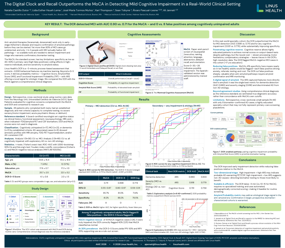
KEY RESULT: The DCR detected MCI with AUC 0.90 vs. 0.71 for the MoCA – and 0 vs. 8 false positives among cognitively unimpaired adults
Background
Anti-amyloid therapies (lecanemab, donanemab) work only in early-stage Alzheimer's disease and require confirmation of amyloid pathology before they can be started. Yet more than 90% of MCI cases go undetected, and only ~1 in 3 people with MCI actually harbor AD pathology — so scalable tools are needed to detect impairment and triage who warrants confirmatory biomarker testing.
The MoCA, the standard screen, has key limitations: specificity as low as 40–50% in primary care (high false positives), ceiling effects in high-reserve individuals, and low sensitivity to amyloid status.
Linus Health's DCR is a ~3-minute, process-based assessment that extracts ~2,000 behavioral features via machine learning. Beyond a 0–5 score, it derives probability metrics — Cognition Score, Amyloid Risk Score (ARS), and Functional Impairment Probability (FIP) — with ARS offering a novel signal for etiologic triage. We compared the DCR with the MoCA against a comprehensive clinical diagnosis.
Methods
Design. Retrospective, cross-sectional study using routine-care data from the Neurology Unit, Universidad Católica de Valencia (UCV), Spain. Patients evaluated for cognitive concerns completed both the MoCA and DCR and consented to research use.
Sample. 49 patients who completed both tests, had an established diagnosis, and met criteria (capacity to complete testing; no severe sensory/motor impairment, acute psychiatric illness, or delirium).
Reference standard. A board-certified neurologist set cognitive status via clinical history, functional assessment, neuropsychology, MRI, and—when indicated—FDG/amyloid PET and CSF biomarkers. DCR and MoCA scores were not used in diagnosis.
Classification. Cognitively unimpaired (n=17), MCI (n=21), or dementia (n=11) by established criteria. AD-associated cases (n=8) showed amnestic profiles with MRI atrophy, FDG-PET hypometabolism, and/or AD-consistent CSF.
Analyses. Analysis 1 (N=38): CU vs. MCI. Analysis 2 (N=49): CU vs. all cognitively impaired, with exploratory AD vs. non-AD etiology.
Statistics. t-tests / Fisher's exact test; ROC-AUC with 1,000-bootstrap 95% CIs and DeLong's test; Youden-index cutoffs; concordance (Cohen's κ); and MoCA-negative rescue analyses (MATLAB R2025b).
Table 1. CU and MCI groups were comparable in age, sex, and education (all p>0.1)
| Characteristic | CU (n=17) | MCI (n=21) |
|---|---|---|
| Age, yrs | 54.6 ± 15.4 | 55.2 ± 18.9 |
| Male, n (%) | 7 (41%) | 12 (57%) |
| Education, yrs | 17.8 ± 0.7 | 17.3 ± 1.0 |
| MoCA | 25.7 ± 2.5 | 22.1 ± 5.2 |
| DCR 0–5 Score | 4.1 ± 0.8 | 2.0 ± 1.5 |
Study Design — Figure 1. Workflow. The UCV cohort was assessed with MoCA and DCR during routine care; comprehensive clinical diagnosis was the reference standard.
Cognitive Assessments
Figure 2. Digital Clock and Recall (DCR): Digital clock drawing test plus 3-word Immediate and Delayed Recall, on an iPad.
| DCR output | What it indicates |
|---|---|
| DCR 0–5 Score | Overall performance (recall + clock) |
| Cognition Score | Probability of cognitive impairment |
| Amyloid Risk Score (ARS) | Probability of elevated brain amyloid |
| FIP | Probability of functional impairment |
MoCA. Paper-and-pencil screen of visuospatial/executive, naming, attention, language, abstraction, delayed recall, and orientation. Score 0–30; <26 indicates possible impairment. The Spanish-validated version was administered by trained staff.
Figure 3. The Montreal Cognitive Assessment (MoCA).
Results
Primary — MCI detection (CU vs. MCI, N=38)
Figure 4. ROC curves for MCI detection. The DCR 0–5 Score (orange) reached AUC 0.898 vs. 0.711 for MoCA (ΔAUC +0.187, p<0.001).
Table 2. DCR vs. MoCA: higher AUC, far higher specificity, fewer false pos.
| MoCA | DCR 0–5 | DCR Cog. | |
|---|---|---|---|
| AUC | 0.711 | 0.898 | 0.774 |
| 95% CI | 0.55–0.87 | 0.80–0.97 | 0.59–0.91 |
| Sensitivity | 85.7% | 81.0% | 71.4% |
| Specificity | 41.2% | 88.2% | 76.5% |
| False pos. (N) | 8 | 0 | 1 |
Among 17 cognitively unimpaired adults, MoCA flagged 8 false positives – the DCR 0–5 Score flagged 0.
MoCA-negative rescue: of 14 MoCA-negative cases (≥26), 5 were truly impaired; the DCR correctly flagged 40% (2/5) that MoCA classified as normal.
At 20% prevalence: the DCR 0–5 Score yields PPV 63% and NPV 95%, supporting use as a rule-out screen.
Secondary — any impairment & etiology (N=49)
Figure 5. ROC curves for any cognitive impairment (CU vs. CI). The DCR 0–5 Score reached AUC 0.931 vs. 0.776 for MoCA.
Table 3. Exploratory analysis (n=8 AD-confirmed). DCR probability metrics outperform MoCA across tasks.
| Clinical task | Best DCR metric | DCR AUC | MoCA AUC |
|---|---|---|---|
| Dementia detection | ARS | 0.880 | 0.833 |
| Severity (MCI vs. dem.) | ARS | 0.818 | 0.779 |
| Etiology (AD vs. non-AD) | Cognition Score | 0.791 | 0.758 |
Figure 6. Exploratory (n=8 AD): ARS rises across CU < MCI < dementia (left) and discriminates AD- from non-AD etiology (right; AUC 0.756).
Discussion
In this real-world specialty cohort, the DCR outperformed the MoCA for MCI detection (AUC 0.898 vs. 0.711) and for any cognitive impairment (0.931 vs. 0.776), while substantially improving specificity.
Penetrating cognitive reserve. Cognitive reserve allows highly educated patients to achieve normal scores on output-based tests despite pathology, but the neural cost — slower processing, motor hesitations, compensatory strategies — leaves traces in the DCR's high-resolution data. The DCR flagged MoCA-negative MCI cases in this cohort (>17 yrs education).
Reducing false positives. MoCA's 41% specificity here means nearly six in ten healthy adults would be flagged—each false positive driving anxiety, referrals, testing, and cost. The DCR cut false positives sharply, valuable when anti-amyloid pathways require amyloid confirmation and MRI monitoring.
A digital signal of amyloid. The ARS captured features more directly tied to amyloid: it was the single best metric for dementia detection (AUC 0.880) and severity staging (0.818) and discriminated AD from non-AD etiology.
Beyond agreement studies. Using comprehensive clinical diagnosis as the reference standard, we estimated true diagnostic accuracy rather than concordance with MoCA's own cutoffs.
Limitations. Retrospective, cross-sectional, small sample (N=49) with only 8 biomarker-confirmed AD cases; a highly educated specialty cohort that may not fully represent primary-care screening populations.
Conclusions
The DCR improved early impairment detection while reducing false positives relative to the MoCA.
Two-dimensional triage. High impairment + high ARS may indicate probable AD warranting PET/CSF; high impairment + low ARS suggests non-AD causes—directing biomarker testing to those most likely to benefit.
Scalable & efficient. The DCR takes ~3 min (vs. 10–15 for MoCA), requires no specialized training, and uses automated, demographically corrected scoring—making it feasible for routine screening.
Amyloid Probability metric may add an etiological triage signal in the anti-amyloid era. Confirmation in larger, prospective, biomarker-characterized cohorts is warranted.
Figure 7. DCR-enabled pathway: pairing cognitive impairment probability with Amyloid Risk Score (ARS) to prioritize biomarker testing.
References
- Nasreddine et al. The MoCA: a brief screening tool for MCI. J Am Geriatr Soc. 2005;53(4):695-9.
- Jannati et al. Digital Clock and Recall is superior to the MMSE for detecting MCI and mild dementia. Alzheimers Res Ther. 2024;16(1):2.
- Gomes-Osman et al. Digital Clock and Recall: a process-driven evolution of the Mini-Cog. Front Hum Neurosci. 2024;18:1337851.
- Jannati et al. Concurrent detection of cognitive impairment and amyloid positivity with a multimodal ML-enabled digital cognitive assessment. Alzheimers Res Ther. 2025;17:261.
Abbreviations: CU/CI, cognitively unimpaired / impaired; MCI, mild cognitive impairment; ARS, Amyloid Risk Score; FIP, Functional Impairment Probability; DCR, Digital Clock & Recall; DCT, Digital Clock Drawing Test (part of DCR); PET/CSF, amyloid-confirmation biomarkers.
Ethics: all participants provided written informed consent for research use.
Disclosures: K. Thompson, S. Tobyne, Á. Pascual-Leone, and A. Jannati are affiliated with Linus Health.
AAIC 2026 • © 2026 Linus Health, Inc. • ajannati@linus.health • linushealth.com
Comments
Email is required but is not shown publicly.
Loading comments...