4,068 posters, 219 videos, 101 topics, 69 sessions, 14,365 authors, 6,128 institutions
ePostersLive by SciGen Technologies S.A. All rights reserved.
Alzheimer's Association International Conference 2026
July 12-15, 2026 | London, United Kingdom, and Online

7355
Dissociating PET-determined Amyloid Positive Vs. Negative Memory Clinic Patients With Linus Digital Assessment Of Cognition (DAC)
Poster Presenter
Authors
Mitchel Kling, Kallie Sweetman, Jacquelyn Nguyen, Amanda Bernardini, Alvaro Pascual-Leone, Rodney Swenson, Christopher Janson, David Libon
Affiliations
Department of Geriatrics and Gerontology, Rowan-Virtua School of Osteopathic Medicine, Stratford NJ USA, Rowan-Virtua School of Osteopathic Medicine, Stratford NJ USA, Department of Psychiatry, Inspira Medical Center, Vineland NJ USA, Department of Neurology, Harvard Medical School, Boston MA USA, Department of Neuroscience, University of North Dakota School of Medicine and Health Sciences, Grand Forks ND USA, Department of Cell Biology, Rowan-Virtua School of Osteopathic Medicine, Stratford NJ USA, Department of Geriatrics and Gerontology, Rowan-Virtua School of Osteopathic Medicine, Stratford NJ USA and Department of Psychology, Rowan University, Glassboro NJ
Part of Topic
Background: PET scans are frequently used to determine amyloid status. The current research sought to
characterize patterns of neuropsychological impairment among memory clinic patients determined to be
amyloid positive versus amyloid negative by PET.
Methods: 79 patients from the New Jersey Institute for Successful Aging underwent neuropsychological
assessment using the Linus Digital Assessment of Cognition (DAC), a brief, 7-minute digitally
administered/scored, protocol measuring verbal episodic memory (6-word Philadelphia repeatable Verbal
Learning Test [PrVLTI, executive abilities (Backward Digit Span Test [BOST], and language ('animal' fluency
test). Blood-based dementia-related biomarkers were obtained with a commercial ATN panel (A~42/A~40
ratio, P-tau181, Nfl). 18F-florbetaben (Neuraceq®) PET scans were used to determine amyloid status.
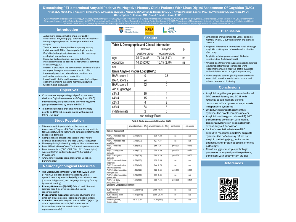
Results: 62 patients (78%) were amyloid positive, and 17 (22%) were amyloid negative. Groups
(female=63.2%) were equated for age/education (age=75.91±4.89; education=15.00±2.66; Table 1). On the
P(r)VLT, the PET amyloid positive group scored lower on immediate free recall, delayed free recall, and
recognition tests conditions (p< 0.032). The PET amyloid positive group also presented with a lower
delayed free recall/immediate free recall savings index (p< 0.001), produced fewer free recall cluster
responses (p< 0.039), and endorsed more semantic recognition foils (p< 0.001). Groups did not differ on
the BOST indices assessing auditory span and verbal working memory. Regarding language, groups did not
differ for output on the 'animal' fluency test. However, on this test the amyloid negative group were more
likely to use semantic strategies ('animal' Association Index) to aid test performance (p< 0.023). When a
stepwise nominal regression analysis using blood-based ATN markers (A~42/ A~40 ratio, P-tau181, Nfl},
was conducted to classify patients, only P-tau181 entered the final model (Wald=4.58, p< 0.032).
Conclusion: The amyloid PET positive group presented with a mixed neuropsychological profile involving
amnesia, degraded semantic functioning, and impaired executive abilities. The amyloid PET negative group
was characterized primarily by executive dysfunction with better semantic abilities. The data reported
above show how a brief digital neuropsychological protocol can effectively characterize neurocognitive
strengths and weaknesses in a pathophysiologically-relevant manner. The imaging and blood-based
biomarker results suggest that multiple neuropathologic substrates could underlie the neuropsychological
problems in the PET amyloid positive group.
Comments
Email is required but is not shown publicly.
Loading comments...