4,068 posters, 219 videos, 101 topics, 69 sessions, 14,365 authors, 6,128 institutions
ePostersLive by SciGen Technologies S.A. All rights reserved.
Alzheimer's Association International Conference 2026
July 12-15, 2026 | London, United Kingdom, and Online

3274
Loss of phasic locus coeruleus activity is associated with increases in tau accumulation and cognitive decline
Poster Presenter
Authors
Prokopis Prokopiou, Aaron Schultz, Maxime Van Egroo, Joost Riphagen, Nicholas Ashton, Shorena Janelidze, Kathryn Papp, David Salat, Dorene Rentz, Kaj Blennow, Oskar Hansson, Henrik Zetterberg, Reisa Sperling, Keith Johnson, Heidi Jacobs
Affiliations
Clinical Memory Research Unit, Department of Clinical Sciences Malmö, Lund University, Lund, Sweden, Athinoula A. Martinos Center for Biomedical Imaging, Department of Radiology, Massachusetts General Hospital, Boston, MA 02129, USA, Department of Psychiatry and Neurochemistry, Institute of Neuroscience and Physiology, The Sahlgrenska Academy, Gothenburg, 431 41 Mölndal, Sweden, Center for Alzheimer Research and Treatment, Department of Neurology, Brigham and Women’s Hospital, Boston, MA, USA, Gordon Center for Medical Imaging, Department of Radiology, Massachusetts General Hospital, Boston, MA, USA
Part of Topic
Loss of phasic activity is associated with increases in tau accumulation and cognitive decline
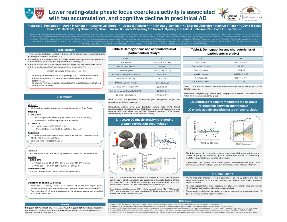
Background: The locus coeruleus (LC), a small brainstem nucleus and the brain’s primary source of norepinephrine, is among the earliest regions harboring tau in Alzheimer’s disease (AD), years before cortical tau involvement. Previous studies in transgenic AD mice overexpressing pretangle tau in the LC indicated that phasic LC activation, characterized by burst-like, high-frequency firing (>15Hz), may protect against LC tau spread and its effects on cognition. We examined whether in-vivo LC phasic activity extracted from resting-state (rs)fMRI data relates to tau accumulation and cognitive decline in asymptomatic older individuals.
Method: We analyzed two cohorts (Table1): (1) 92 Harvard Aging Brain Study participants (mean age=74.99±9.0 years, female=61%) who underwent rsfMRI, [18F]-FTP(tau)-PET, and longitudinal cognitive testing (follow-up=5.37±1.88 years). (2) 68 individuals (age=60.21±12.01 years, female=50%) who underwent (7T)-rsfMRI, plasma biomarker assessment (GFAP, p-tau217/181/231), and longitudinal cognitive testing (follow-up=3.02±1.1 years). Phasic-like LC activity was defined as BOLD-fMRI signal peaks detected with an in-house minimum-entropy–based event-detection algorithm, optimized for concurrent entorhinal (EC) signal integration, given that the EC is the earliest cortical site of tau pathology and LC–EC decoupling may indicate LC tau spread. Linear regression and mixed-effects analyses associated phasic LC activity with tau and cognitive decline, adjusted for age, sex, and education.
Result: (1) Lower phasic LC activity was associated with greater EC tau deposition (Fig.2A-B), faster inferior-temporal cortical tau accumulation (Fig.2C), and steeper decline in PACC (Fig.2D). (2) No associations between phasic LC activity and plasma tau or GFAP were observed (Fig.3A). However, lower LC activity was associated with higher GFAP-related p-tau231 levels (Fig.3B). Furthermore, we found no significant association between phasic LC activity and the PACC. Post-hoc analyses of PACC subtests revealed that lower LC activity was associated with steeper decline in verbal fluency, particularly at elevated p-tau231 and GFAP levels (Fig.3C).
Conclusion: Our findings, consistent with prior animal and task-fMRI evidence, suggest that lower resting-state phasic LC activity may contribute to tau progression and memory decline in asymptomatic older adults, with astrogliosis representing a potential mechanistic pathway. Our findings could inform early targeted interventions aimed at supporting optimal LC phasic activity, with the potential to prevent or delay disease progression.
Comments
Email is required but is not shown publicly.
Loading comments...