4,068 posters, 219 videos, 101 topics, 69 sessions, 14,365 authors, 6,128 institutions
ePostersLive by SciGen Technologies S.A. All rights reserved.
Alzheimer's Association International Conference 2026
July 12-15, 2026 | London, United Kingdom, and Online

5536
Extracranial Carotid Artery Disease Is Associated With Reduced Ipsilateral Brain Volume Through Reductions In Cortical Surface Area
Poster Presenter
Authors
Haley Wiskoski, Summan Zahra, Juan Arias, Scott French, Kevin Johnson, Raza Mushtaq, Gene Alexander, Theodore Trouard, Craig Weinkauf
Affiliations
Department of Neuroradiology, Barrow Neurological Institute, Phoenix, AZ, USA, Department of Radiology and Imaging Sciences, University of Arizona, Tucson, AZ, USA, BIO5 Research Institute, University of Arizona, Tucson, AZ, USA; Evelyn F. McKnight Brain Institute, Tucson, AZ, USA; Departments of Psychology and Psychiatry and Neuroscience and Physiological Sciences Graduate Interdisciplinary Programs, University of Arizona, Tucson, AZ, USA; Arizona Alzheimer’s Consortium, Phoenix, AZ, USA, Department of Biomedical Engineering, University of Arizona, Tucson, AZ, USA; Department of Radiology and Imaging Sciences, University of Arizona, Tucson, AZ, USA; BIO5 Research Institute, University of Arizona, Tucson, AZ, USA; Evelyn F. McKnight Brain Institute, Tucson, AZ, USA, Department of Biomedical Engineering, University of Arizona, Tucson, AZ, USA; Department of Surgery, University of Arizona, Tucson, AZ, USA; Department of Neuroscience, University of Arizona, Tucson, AZ, USA, Department of Biomedical Engineering, University of Arizona, Tucson, AZ, USA; Department of Surgery, University of Arizona, Tucson, AZ, USA, Department of Surgery, University of Arizona, Tucson, AZ, USA
Part of Topic
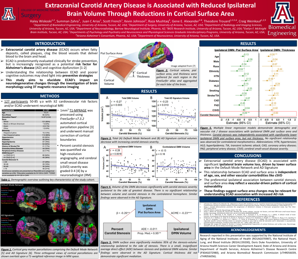
Background: Asymptomatic extracranial carotid artery disease (aECAD) is a prevalent large-vessel cerebrovascular disease (CVD) and an emerging risk factor for Alzheimer’s disease (AD) and related dementias. While cortical thinning is well described in aging and AD, it remains unclear how CVD, including aECAD and cerebral small vessel disease (cSVD), alters the morphometric components of volume—surface area (SA) and thickness—which possess distinct developmental and cognitive associations. Here, we assess aECAD-related morphometric alterations within an AD-vulnerable network.
Methods: 127 adults (50-85yrs) from the Carotids and Minds prospective cohort with ≥2 cardiovascular risk factors, with/without aECAD and cSVD, underwent brain MRI including 1mm3 T1-MPRAGE. Cortical parcellation was performed using FreeSurfer v7.3.2 with manual editing. Percent carotid stenosis was quantified via high-resolution angiography and cSVD severity was graded (0-4) by a neuroradiologist. Default Mode Network (DMN) morphometrics were gathered ipsilateral and contralateral to maximum stenosis. Regressions tested hemispheric morphometric associations, and causal mediation analyses evaluated direct and indirect effects of stenosis on volume through SA and thickness.
Results: aECAD severity was associated with lower ipsilateral DMN volume (r=-0.28, p=0.0016), but not contralateral (r=-0.13, p=0.16). Contralateral white-matter (WM) SA, pial SA, and thickness were not significantly related to stenosis. In multivariable models adjusting for age, sex, CVD and risk factors, percent stenosis remained an independent predictor of reduced ipsilateral DMN volume (p=0.021) and SA (WM: p=0.004; pial: p=0.007), but not thickness (p=0.89). cSVD showed no significant association with these metrics. Mediation analysis showed ipsilateral pial SA significantly mediated the stenosis-volume relationship (ACME=-0.23, p<0.001) with a small direct effect (ADE=-0.11) accounting for 95% of the total effect (p<0.01), whereas thickness mediated ~5% (ACME=-0.013, ADE=-0.22).
Conclusion: aECAD was associated with lateralized structural vulnerability in the DMN. cSVD was found to have no significant effect on morphometry. The strong mediation of the stenosis-volume relationship by SA, not thickness, suggests a vascular-specific remodeling process distinct from the canonical thickness-dominant atrophy emphasized in aging and AD. These findings position cortical SA as a potentially sensitive marker of vascular disease progression with potential to improve detection of vascular contributions to AD-related brain changes.
Comments
Email is required but is not shown publicly.
Loading comments...