4,068 posters, 219 videos, 101 topics, 69 sessions, 14,365 authors, 6,128 institutions
ePostersLive by SciGen Technologies S.A. All rights reserved.
Alzheimer's Association International Conference 2026
July 12-15, 2026 | London, United Kingdom, and Online

10988
High-resolution Computational Histology Resolves Ongoing Amyloid-beta Accumulation Beyond The Neuroimaging Plateau
Poster Presenter
Authors
Part of Topic
BACKGROUND: Current neuroimaging frameworks,
including the NIA-AA 2024 Revised Criteria, describe a
"plateau" in amyloid-beta (A-Beta) burden during the
symptomatic stages of Alzheimer's disease. While PET
imaging provides a vital macroscopic view, its resolution may
mask ongoing microscopic accumulation. We developed a
computational pathology pipeline to segment and classify
various A-Beta burden, using high-resolution histological
quantification to unmask continuous pathological growth
across Thal phases.
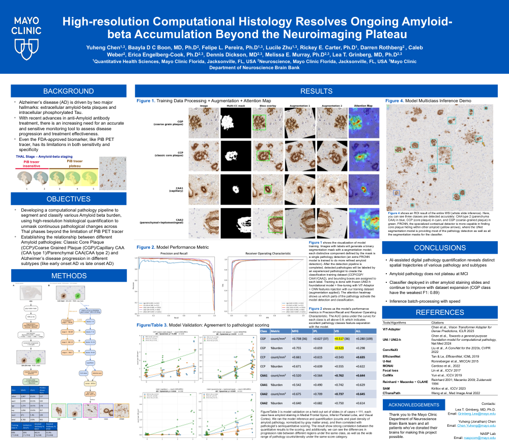
METHODS: We assembled a corpus of 7,940
neuropathologist-validated amyloid lesion instances
sampled from 321 whole-slide images stained for Aβ (6E10
and 6F3D), spanning five neuroanatomical regions (middle
frontal, inferior parietal, visual, parahippocampal, and motor
cortex). A two-stage pipeline was developed: (1) an
EfficientNet-B4 U-Net segments Aβ-immunoreactive deposits
to quantify pixel-level total burden, and (2) a classifier built
on UNI2-h, a pathology foundation model, adapted with a
trainable convolutional branch through gated cross-attention
(ViT-Adapter) and deployed as a dual-specialist, five-fold
cross-validated ensemble, assigns each deposit to one of five
classes: coarse-grained plaque (CGP), classic cored plaque
(CCP), capillary (CAA1) and parenchymal/leptomeningeal
(CAA2) cerebral amyloid angiopathy, or non-specific/other.
Internal performance was assessed by out-of-fold (OOF)
cross-validation; external validity was assessed by correlating
model-derived per-region deposit counts with independent
neuropathologist semi-quantitative scores.
RESULTS: The classifier achieved an OOF macro-F1 of
0.92 (per-class F1: 0.91 CGP, 0.90 CCP, 0.92 CAA1, 0.95 CAA2,
0.91 non-specific). Model-derived per-region deposit counts
correlated with independent neuropathologist semiquantitative
scores, with strongest agreement in the region
where each morphology predominates: CGP in middle
frontal cortex (Spearman ρ=0.755), CAA2 across all regions
(ρ=0.64–0.75), CAA1 in visual cortex (ρ=0.742), and CCP
across regions (ρ=0.55–0.67); all p<0.005.
CONCLUSIONS: A pathology foundation-model
classifier, coupled to deep segmentation and refined through
iterative neuropathologist quality control, enables accurate,
reproducible, morphology-resolved quantification of cerebral
Aβ pathology across regions, with high concordance to expert
scoring. This provides a scalable, higher-fidelity tool for
precision neuropathological staging and validating antiamyloid
therapeutic response.
Comments
Email is required but is not shown publicly.
Loading comments...