4,068 posters, 219 videos, 101 topics, 69 sessions, 14,365 authors, 6,128 institutions
ePostersLive by SciGen Technologies S.A. All rights reserved.
Alzheimer's Association International Conference 2026
July 12-15, 2026 | London, United Kingdom, and Online

4385
Predicting Amyloid PET Positivity Using Brain Perfusion Single Photon Emission Computed Tomography
Poster Presenter
Authors
Affiliations
Part of Topic
Background
Brain perfusion single-photon emission computed tomography (SPECT) with the easy Z-score Imaging System (eZIS) has emerged as a practical quantitative tool for detecting regional cerebral blood flow abnormalities associated with early Alzheimer’s disease. Given the growing need for accessible biomarkers that can predict amyloid pathology, evaluating the diagnostic performance of eZIS carries important clinical implications.
Methods
The present study retrospectively analyzed patients with mild cognitive impairment and dementia due to Alzheimer’s disease, who underwent both 99mTc-ECD SPECT with eZIS quantification and amyloid Positron Emission Tomography (PET) using 18F-florbetaben. Three eZIS parameters in the target regions (precuneus/posterior cingulate and parietal cortices) were analyzed: severity (degree of hypoperfusion), extent (% of voxels exceeding Z-score threshold), and ratio (rCBF reduction in the volume of interest compared with that of the whole brain). Amyloid PET positivity was determined by visual assessment using the BAPL score. Patients were classified as eZIS-positive or -negative based on predefined cutoff values. In addition, receiver operating characteristic (ROC) curve analyses were performed to evaluate the ability of these parameters to predict amyloid positivity and to identify the optimal discriminative approach.
Results
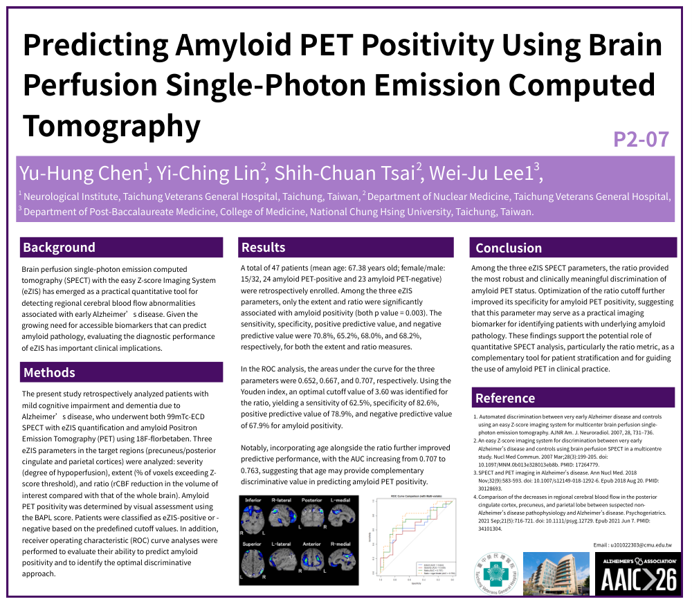
A total of 47 patients (mean age: 67.38 years old; female/male: 15/32, 24 amyloid PET-positive and 23 amyloid PET-negative) were retrospectively enrolled. Among the three eZIS parameters, only the extent and ratio were significantly associated with amyloid positivity (both p value = 0.003). The sensitivity, specificity, positive predictive value, and negative predictive value were 70.8%, 65.2%, 68.0%, and 68.2%, respectively, for both the extent and ratio measures.
In the ROC analysis, the areas under the curve for the three parameters were 0.652, 0.667, and 0.707, respectively. Using the Youden index, an optimal cutoff value of 3.60 was identified for the ratio, yielding a sensitivity of 62.5%, specificity of 82.6%, positive predictive value of 78.9%, and negative predictive value of 67.9% for amyloid positivity.
Notably, incorporating age alongside the ratio further improved predictive performance, with the AUC increasing from 0.707 to 0.763, suggesting that age may provide complementary discriminative value in predicting amyloid PET positivity.
Conclusion
Among the three eZIS SPECT parameters, the ratio provided the most robust and clinically meaningful discrimination of amyloid PET status. Optimization of the ratio cutoff further improved its specificity for amyloid PET positivity, suggesting that this parameter may serve as a practical imaging biomarker for identifying patients with underlying amyloid pathology. These findings support the potential role of quantitative SPECT analysis, particularly the ratio metric, as a complementary tool for patient stratification and for guiding the use of amyloid PET in clinical practice.
Comments
Email is required but is not shown publicly.
Loading comments...