4,068 posters, 219 videos, 101 topics, 69 sessions, 14,365 authors, 6,128 institutions
ePostersLive by SciGen Technologies S.A. All rights reserved.
Alzheimer's Association International Conference 2026
July 12-15, 2026 | London, United Kingdom, and Online

1445
Understanding The Impact Of Anti-aί Immunotherapy On Myeloarchitectural Organization
Poster Presenter
Authors
Part of Topic
Background:
Anti-amyloid immunotherapies are designed to target Aβ plaques. These second-generation monoclonal antibodies (mAbs) instigate transient immune responses by recruiting the brain’s own immune system leading to phagocytosis of cerebral Aβ. During the active treatment phase, these mAbs can trigger oedema/effusion and microhemorrhages, termed amyloid related imaging abnormalities (ARIA). Accelerated brain volume loss has also been reported as one of the major downstream side effects. The pathological mechanisms and long-term implication of “volume loss” on brain health are still understudied. Since microstructural alterations precede macro-structural changes, characterizing myeloarchitectural remodeling may identify imaging biomarkers sensitive to upstream immunotherapy effects. The aim of the current study is to map myeloarchitectural alterations in Tg2576 mice following 12-week immunotherapy using ultra-high resolution quantitative transverse relaxation (qT2) and myelin water fraction (MWF) imaging.
Method:
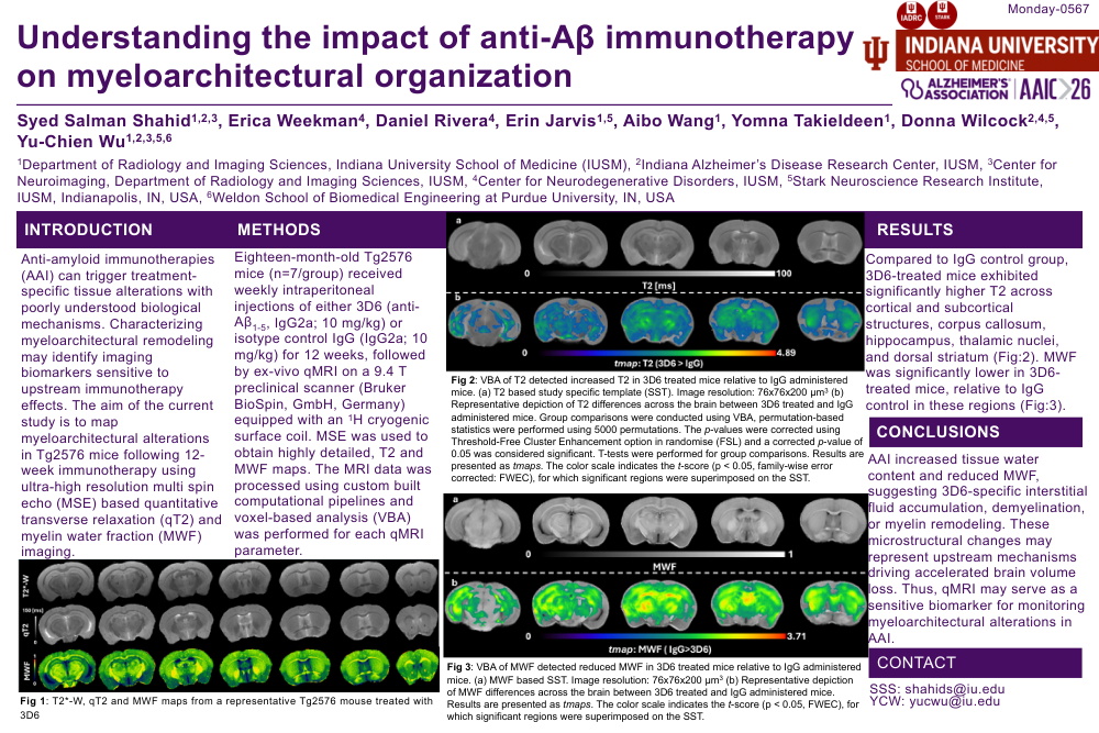
Eighteen-month-old Tg2576 mice received weekly intraperitoneal injections of either 3D6 (anti-Aβ1-5, IgG2a; 10 mg/kg; n=7) or isotype control IgG (IgG2a; 10 mg/kg; n=7) for 12 weeks. Mice were sacrificed 3 days post final injection and brains harvested and fixed for ultra-high resolution ex-vivo qMRI on a 9.4 T preclinical scanner equipped with an 1H cryogenic surface coil. Multi-echo spin echo imaging was used to obtain highly detailed, transverse relaxation (T2) maps and MWF maps. The MRI data was processed using custom built computational pipelines and voxel-based analysis (VBA) was performed for each qMRI parameter (Figures 1 and 2).
Results:
T2 and MWF based VBA showed widespread differences between groups. Compared to IgG control group, 3D6-treated mice exhibited significantly higher T2 across cortical and subcortical structures, corpus callosum, hippocampus, thalamic nuclei, and dorsal striatum (Figure 1), indicative of increased interstitial water content. MWF was significantly lower in 3D6-treated mice, relative to IgG control in these regions, indicating immunotherapy induced myeloarchitectural remodeling (Figure 2).
Conclusion:
Anti-Aβ immunotherapy with 3D6 induces widespread elevations in tissue water content and reductions in MWF, suggesting treatment-associated demyelination or myelin remodeling. These microstructural changes may represent upstream mechanisms contributing to the accelerated brain volume loss observed clinically. qT2 and MWF imaging offer sensitive biomarkers for monitoring myeloarchitectural effects of anti-amyloid therapies.
Comments
Email is required but is not shown publicly.
Loading comments...