4,068 posters, 219 videos, 101 topics, 69 sessions, 14,365 authors, 6,128 institutions
ePostersLive by SciGen Technologies S.A. All rights reserved.
Alzheimer's Association International Conference 2026
July 12-15, 2026 | London, United Kingdom, and Online

9035
Association Between Plasma GFAP And FDG PET Radiomic Features In The Medial Temporal Lobe: A Pilot Investigation Using The Adni Cohort
Part of Topic
TITLE: Association Between Plasma GFAP And FDG PET Radiomic Features In The Medial Temporal Lobe: A Pilot Investigation Using The Adni Cohort
AUTHORS: Nese Rihani, Gokay Mutlu, Ramin Rasi, Ozgur Koray Sahingoz
POSTER NUMBER: P2-06
INTRODUCTION:Plasma glial fibrillary acidic protein (GFAP) is an established blood biomarker for astrogliosis and the early Alzheimer's disease (AD) pathology. Although FDG-PET is conventionally used to assess neurodegeneration through regional glucose metabolism, radiomic analysis allows the consideration of imaging features. This study investigated whether FDG-PET radiomic features in Alzheimer’s-vulnerable medial temporal lobe (MTL) regions reflect systemic astroglial activation across the AD spectrum.
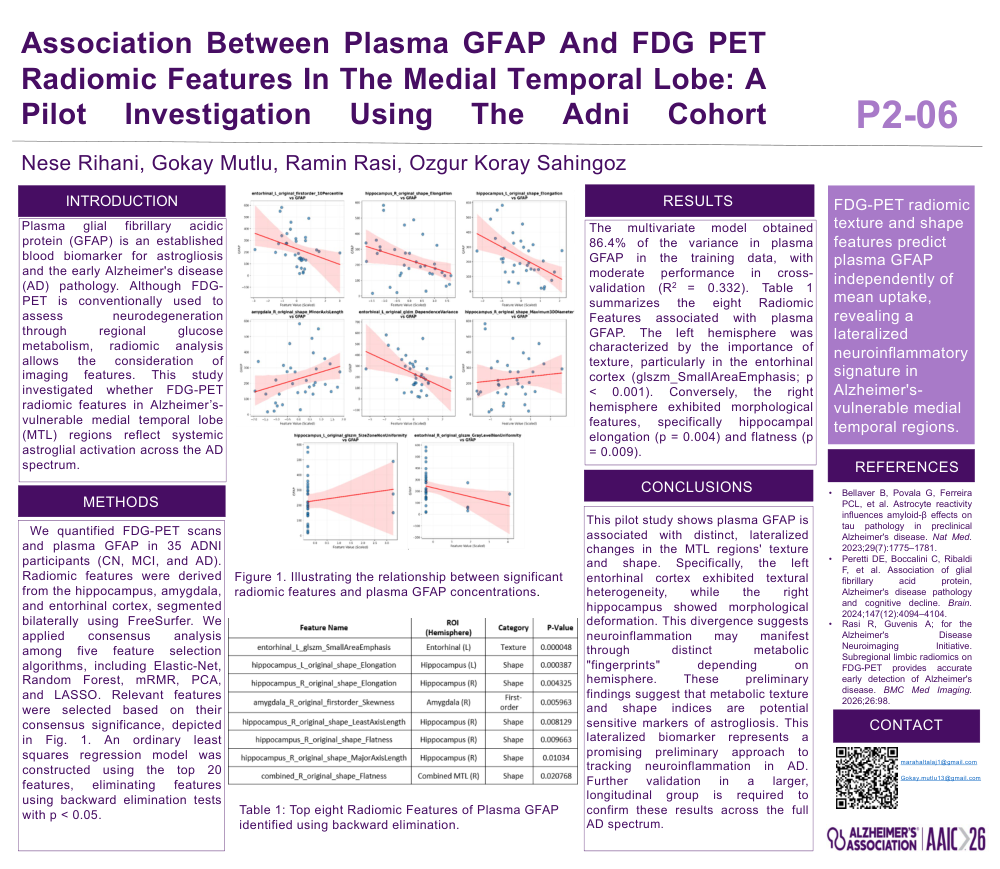
METHODS: We quantified FDG-PET scans and plasma GFAP in 35 ADNI participants (CN, MCI, and AD). Radiomic features were derived from the hippocampus, amygdala, and entorhinal cortex, segmented bilaterally using FreeSurfer. We applied consensus analysis among five feature selection algorithms, including Elastic-Net, Random Forest, mRMR, PCA, and LASSO. Relevant features were selected based on their consensus significance, depicted in Fig. 1. An ordinary least squares regression model was constructed using the top 20 features, eliminating features using backward elimination tests with p < 0.05.
RESULTS:The multivariate model obtained 86.4% of the variance in plasma GFAP in the training data, with moderate performance in cross-validation (R2 = 0.332). Table 1 summarizes the eight Radiomic Features associated with plasma GFAP. The left hemisphere was characterized by the importance of texture, particularly in the entorhinal cortex (glszm_SmallAreaEmphasis; p < 0.001). Conversely, the right hemisphere exhibited morphological features, specifically hippocampal elongation (p = 0.004) and flatness (p = 0.009).
CONCLUSİON:This pilot study shows plasma GFAP is associated with distinct, lateralized changes in the MTL regions' texture and shape. Specifically, the left entorhinal cortex exhibited textural heterogeneity, while the right hippocampus showed morphological deformation. This divergence suggests neuroinflammation may manifest through distinct metabolic "fingerprints" depending on hemisphere. These preliminary findings suggest that metabolic texture and shape indices are potential sensitive markers of astrogliosis. This lateralized biomarker represents a promising preliminary approach to tracking neuroinflammation in AD. Further validation in a larger, longitudinal group is required to confirm these results across the full AD spectrum.
Comments
Email is required but is not shown publicly.
Loading comments...