4,068 posters, 219 videos, 101 topics, 69 sessions, 14,365 authors, 6,128 institutions
ePostersLive by SciGen Technologies S.A. All rights reserved.
Alzheimer's Association International Conference 2026
July 12-15, 2026 | London, United Kingdom, and Online

9055
Associations Of Cerebrospinal Fluid Biomarkers With Hippocampal Volume In A Hispanic Cohort
Poster Presenter
Authors
Alcibiades E Villarreal, Anna V Díaz-Ramírez, Josefina Fletcher, Aaron Benzadon, Pahola Araujo, Ricardo Williams, Rainier Rodríguez, Nelson Novarro, David Dondis, David Anguizola, Judy Mendoza, Verónica Billingslea, Olmedo Almanza, Karen Courville, Sassil Pitti, Héctor Torres, Lizetha Quintero, Dámaso Díaz, Laura Cerrud, Bruno Hammerschlag, Alexis Palacios, Andrés Bernales, Ariel Saldaña, Fernando Sucre, Diana Oviedo, Sofía Rodríguez-Araña, Giselle Rangel, Carolina E Rodríguez, María B Carreira, Gabrielle B Britton
Affiliations
Instituto de Investigaciones Cientificas y Servicios de Alta Tecnologia, Servicio de Neurología, CIDELAS-CSS, Servicio de Neurología, CIDELAS CSS, Hospital Dr. Gustavo Nelson Collado, CSS, Hospiten Paitilla, Caja de Seguro Social, Centro de Vacunación Internacional Cevaxin
Part of Topic
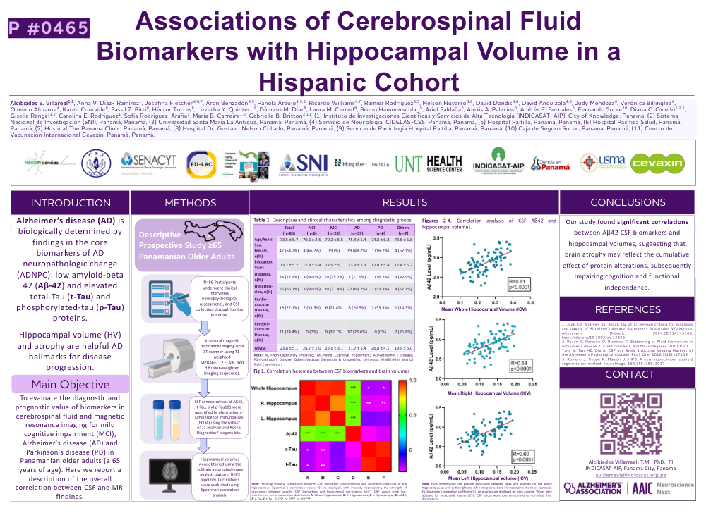
Title: Associations of Cerebrospinal Fluid Biomarkers with Hippocampal Volume in a Hispanic Cohort
Introduction: Alzheimer’s disease (AD) is biologically determined by findings in the core biomarkers of AD neuropathologic change (ADNPC): low amyloid-beta 42 (Aβ-42) and elevated total-Tau (t-Tau) and phosphorylated-tau (p-Tau) proteins. Hippocampal volume (HV) and atrophy are helpful AD hallmarks for disease progression.
Main Objective: To evaluate the diagnostic and prognostic value of biomarkers in cerebrospinal fluid and magnetic resonance imaging for mild cognitive impairment (MCI), Alzheimer's disease (AD) and Parkinson's disease (PD) in Panamanian older adults (≥ 65 years of age). Here we report a description of the overall correlation between CSF and MRI findings in a panamenian cohort of older adults.
Methods: Descriptive and prospective study. Participants underwent clinical interviews, cognitive testing, structural magnetic resonances in a 3T scanner, and lumbar punctures. Sagittal T1 magnetic resonance imaging was acquired using MPRAGE, T2 FLAIR, and diffusion-weighted imaging sequences. Hippocampal volumes were obtained through the volBrain automated image analysis software. Quantification of Aβ-42, t-Tau, p-Tau181 proteins in CSF were determined by ECLIA methodology, with the cobas® e411 analyzer (Roche Diagnostics®) and the corresponding reagent kits ([Ref. 08821909190], [Ref. 07356994 190], [Ref. 08846693190]). Correlations were analyzed with Spearman correlation test.
Conclusion: Our study found significant correlations between Aβ42 CSF biomarkers and hippocampal volumes, suggesting that brain atrophy may reflect the cumulative effect of protein alterations, subsequently impairing cognition and functional independence
Comments
Email is required but is not shown publicly.
Loading comments...