4,068 posters, 219 videos, 101 topics, 69 sessions, 14,365 authors, 6,128 institutions
ePostersLive by SciGen Technologies S.A. All rights reserved.
Alzheimer's Association International Conference 2026
July 12-15, 2026 | London, United Kingdom, and Online

9888
Diagnostic Performance Of Plasma Gfap In Preclinical Stages Of Alzheimer's Disease Using The Lumipulse Platform
Poster Presenter
Authors
Affiliations
Part of Topic
Diagnostic performance of plasma GFAP in preclinical stages of Alzheimer's disease using the Lumipulse platform
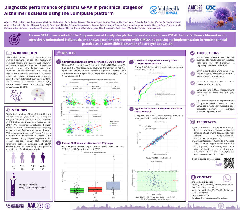
Background: Plasma glial fibrillary acidic protein (GFAP) is a promising biomarker of astrocytic reactivity in preclinical Alzheimer´s Disease (AD). However, most evidence to date comes from ultrasensitive research assays, with limited data from automated clinical platforms. We aimed to evaluate the diagnostic performance of plasma GFAP in cognitively unimpaired (CU) individuals using the fully automated Lumipulse platform, and to assess its concordance with a highly sensitive research-based immunoassay, Single Molecule Array (SIMOA).
Methods: Plasma GFAP, and cerebrospinal fluid (CSF) amyloid-β (Aβ)42/40, phosphorylated tau (p-tau) 181, total tau (t-tau), and neurofilament light chain (NfL) were analyzed in 204 CU participants using the Lumipulse G600II platform. In a subset of 143 individuals, it was also measured with SIMOA. We examined correlations between plasma GFAP and CSF biomarkers, after adjusting for age, sex, and ApoE ε4, and compared plasma GFAP concentrations across AT groups. The ability of plasma GFAP to discriminate amyloid status was assessed using logistic regression and receiver operating curve (ROC) analyses. Agreement between Lumipulse and SIMOA techniques was evaluated using Passing-Bablok regression and Bland-Altman analysis.
Results: Plasma GFAP correlated with Aβ42 (r=-0.24; p-value<0.001) and Aβ42/Aβ40 (Rho=-0.24; p-value<0.001) (Fig. 1). Plasma GFAP concentrations were higher in A+ compared with A− subjects (64.7 vs. 47.6 pg/mL; p=0.001), and in T+ compared with T− (64.3 vs. 48.5 pg/mL; p=0.005). A+T+ subjects showed higher plasma GFAP levels than A-T- (difference=22.2 pg/mL; p<0.0001; Cohen’s d=0.81). This difference remained significant after adjustment for covariates (estimate=14.5 pg/mL; p=0.005) (Fig. 2). Plasma GFAP discriminated amyloid status (A+ vs. A- individuals) with an AUC of 0.67 (95%CI: 0.59-0.76). Lumipulse and SIMOA measurements showed a strong correlation (r=0.96; p<0.001), and good agreement (97% within Bland-Altman limits; slight proportional bias in Passing-Bablok regression) (Fig. 3).
Conclusion: Plasma GFAP measured with the fully automated Lumipulse platform is a reliable and accessible tool for assessing astrocyte activation in preclinical AD. Although its individual diagnostic performance is moderate for detecting amyloid pathology in CU individuals, its relationship with CSF biomarkers and concordance with SIMOA support its clinical utility.
Comments
Email is required but is not shown publicly.
Loading comments...