4,068 posters, 219 videos, 101 topics, 69 sessions, 14,365 authors, 6,128 institutions
ePostersLive by SciGen Technologies S.A. All rights reserved.
Alzheimer's Association International Conference 2026
July 12-15, 2026 | London, United Kingdom, and Online

8797
Perinatal Lead Exposure Associated With Increased ί-amyloid In Aged Murine Brain
Poster Presenter
Authors
Rachel Morgan, Evelyn Matei, Ingrid Bergin, Tomoko Ishikawa, Junru Pan, Katelyn Polemi, Dana Dolinoy, Kelly Bakulski, Justin Colacino
Affiliations
Part of Topic
Perinatal Lead Exposure Associated with Increased β-Amyloid in Aged Murine Brain
RK Morgan, EK Matei, IL Bergin, T Ishikawa, J Pan, KM Polemi, DC Dolinoy, KM Bakulski, JA Colacino
Background
Exposures to neurotoxicants, including lead (Pb), during development are suspected contributors to dementia. Pathological characteristics of dementia include β-amyloid plaques, which result from the aggregation of β-amyloid peptides and contribute to amyloid angiopathy and neuroinflammation. Here, we assess the impact of developmental Pb exposure on β-amyloid.
Methods
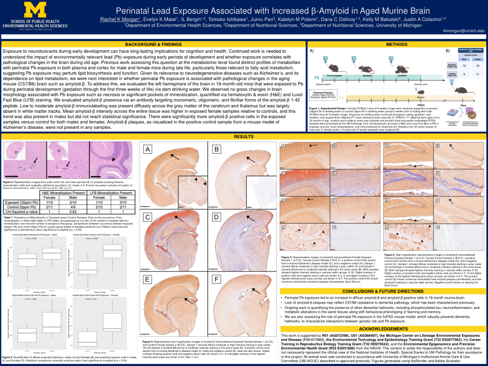
Female C57/B6 mice 6 to 8 weeks of age were exposed to 32 ppm Pb or control water from two weeks prior to mating through offspring weaning, resulting in human-relevant dam blood Pb levels (10 µg/dL) . Offspring were aged to 18 months of age. The left-brain hemisphere was formalin fixed and paraffin embedded (Pb female = 10, Pb male = 10, control female = 10, control male = 11). Sections were stained with hematoxylin and eosin, Luxol Fast blue, or an antibody targeting multiple forms of β-amyloid 1-42. Cells were segmented and staining intensity assessed using QuPath. Differences in diffuse signal, positive cells per tissue area, and mineralization were calculated using a two-sided t-test (significance at p < 0.05) comparing exposed and control, stratified by sex.
Results
β-amyloid plaques were not present in any samples. Low to moderate β-amyloid immunolabeling was present diffusely across the cerebrum and thalamus and more intensely within capillary walls and mineralized thalamic foci. Mean β-amyloid intensity was higher in Pb female samples relative to control (fold change, FC: 1.26, p = 0.004). This trend was also present in male tissues (FC: 1.12) but was not statistically significant (p = 0.095). Exposed females had, on average, 320 β-amyloid positive cells per tissue versus 166 in female control (FC: 1.92, p = 0.011). Exposed males had, on average, 469 β-amyloid positive cells per total tissue slide versus 258 in male controls (FC: 1.82, p = 0.032).
Conclusion
Lack of β-amyloid plaques may reflect C57/B6 resistance to dementia pathology. Perinatal Pb exposure led to an increase in diffuse β-amyloid and β-amyloid-positive cells in aged murine brain. Ongoing work is quantifying the presence of other dementia hallmarks in the same tissues in C57/B6 and 5xFAD mice, which robustly present dementia hallmarks, to characterize interactions between genetic risk and exposure.
Comments
Email is required but is not shown publicly.
Loading comments...