4,068 posters, 219 videos, 101 topics, 69 sessions, 14,365 authors, 6,128 institutions
ePostersLive by SciGen Technologies S.A. All rights reserved.
Alzheimer's Association International Conference 2026
July 12-15, 2026 | London, United Kingdom, and Online

2121
A Preliminary Study Of Variation In Heavy Metal Concentrations Among Brain Donors With And Without Adrd Neuropathology
Poster Presenter
Authors
Affiliations
University of Wisconsin School of Medicine and Public Health
Part of Topic
Background:
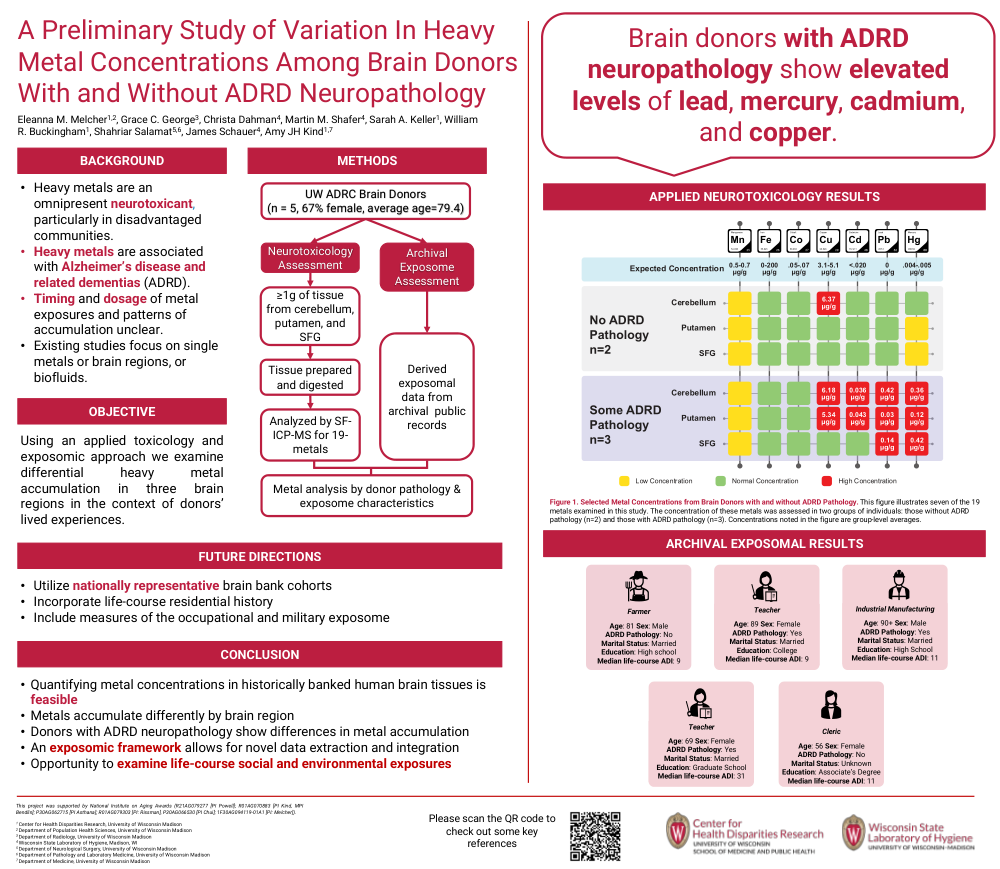
Using an applied toxicology and exposomic approach we examine differential heavy metal accumulation in three brain regions in the context of donors’ lived experiences.
Methods:
We examined the brains of five brain donors (67% female, average age=79.4, average years banked=15, Table 1), using standardized digestion methods based on Krynitsky (1987), in the Trace Elements Clean Lab at Wisconsin State Laboratory of Hygiene. Tissue samples were placed into acid-cleaned and pre-weighed HotBlock digestion tubes and dissolved in concentrated acid. Sample digests were then analyzed by sector field inductively coupled plasma mass spectrometry based on EPA Method 200.8. Analysis of various isotopes were optimized per element to reduce and monitor isobaric interferences. A pre-selected 19 metal panel (Table 2) was examined across three brain regions (superior frontal gyrus (SFG), cerebellum (C), and putamen (P)) to determine high impact regions of interest and to test the feasibility of analysis for future investigations. For our exploratory analyses, we compared tissue concentrations: those with some ADRD pathology and those without.
Results:
We observed higher than expected metal concentration for lead (>0 µg/g, SFG = .14, P=.02, C=.21), mercury (>.005 µg/g, SFG = .21, P=.06, C=.02), cadmium (>.02 µg/g, SFG = .02, P=.03, C=.03), and copper (>5.1 µg/g, SFG = 4.3, P=5.2, C=6.3), especially in the cerebellum and putamen. The additional metals assessed were either below the limit of quantification or within the expected concentrations. In the exploratory analyses, we observed that those with some ADRD pathology had higher than expected metal concentration compared to non-ADRD pathology donors which were within the expected range.
Future Directions:
Conclusion:
Comments
Email is required but is not shown publicly.
Loading comments...