4,068 posters, 219 videos, 101 topics, 69 sessions, 14,365 authors, 6,128 institutions
ePostersLive by SciGen Technologies S.A. All rights reserved.
Alzheimer's Association International Conference 2026
July 12-15, 2026 | London, United Kingdom, and Online

10935
A Night Sky Within The Brain
Part of Topic
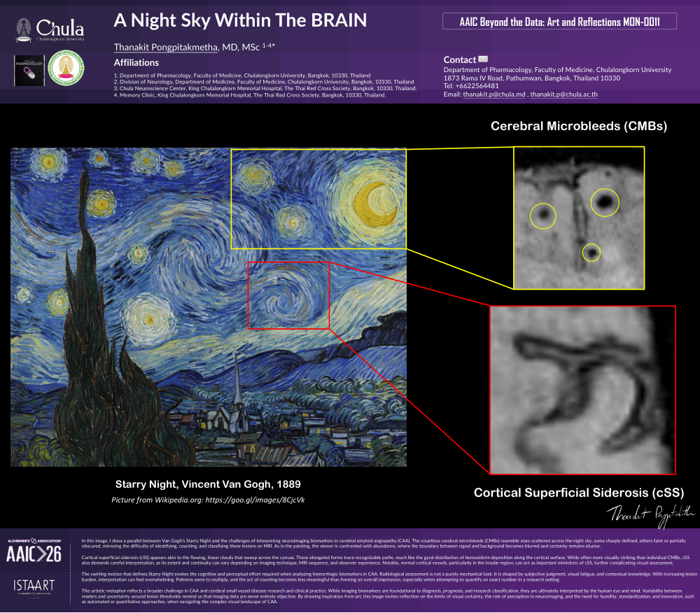
In this image, I draw a parallel between Van Gogh’s Starry Night and the challenges of interpreting neuroimaging biomarkers in cerebral amyloid angiopathy (CAA). The countless cerebral microbleeds (CMBs) resemble stars scattered across the night sky, some sharply defined, others faint or partially obscured, mirroring the difficulty of identifying, counting, and classifying these lesions on MRI. As in the painting, the viewer is confronted with abundance, where the boundary between signal and background becomes blurred and certainty remains elusive.
Cortical superficial siderosis (cSS) appears akin to the flowing, linear clouds that sweep across the canvas. These elongated forms trace recognizable paths, much like the gyral distribution of hemosiderin deposition along the cortical surface. While often more visually striking than individual CMBs, cSS also demands careful interpretation, as its extent and continuity can vary depending on imaging technique, MRI sequence, and observer experience. Notably, normal cortical vessels, particularly in the insular region, can act as important mimickers of cSS, further complicating visual assessment.
The swirling motion that defines Starry Night evokes the cognitive and perceptual effort required when analyzing hemorrhagic biomarkers in CAA. Radiological assessment is not a purely mechanical task. It is shaped by subjective judgment, visual fatigue, and contextual knowledge. With increasing lesion burden, interpretation can feel overwhelming. Patterns seem to multiply, and the act of counting becomes less meaningful than forming an overall impression, especially when attempting to quantify an exact number in a research setting.
This artistic metaphor reflects a broader challenge in CAA and cerebral small vessel disease research and clinical practice. While imaging biomarkers are foundational to diagnosis, prognosis, and research classification, they are ultimately interpreted by the human eye and mind. Variability between readers and uncertainty around lesion thresholds remind us that imaging data are never entirely objective. By drawing inspiration from art, this image invites reflection on the limits of visual certainty, the role of perception in neuroimaging, and the need for humility, standardization, and innovation, such as automated or quantitative approaches, when navigating the complex visual landscape of CAA.
Comments
Email is required but is not shown publicly.
Loading comments...