4,068 posters, 219 videos, 101 topics, 69 sessions, 14,365 authors, 6,128 institutions
ePostersLive by SciGen Technologies S.A. All rights reserved.
Alzheimer's Association International Conference 2026
July 12-15, 2026 | London, United Kingdom, and Online

10645
Arterial Territory Topography Of White Matter Lesions Differentiates Vascular And Neurodegenerative Cohorts
Poster Presenter
Authors
Affiliations
Part of Topic
Arterial Territory Mapping Identifies Diagnosis-Specific White Matter Lesion Topography Across Neurodegenerative and Vascular Syndromes
Background
White matter lesions are a neuroimaging marker commonly associated with cerebral small vessel disease and are linked to cognitive decline and dementia risk. However, it remains unclear whether vascular and neurodegenerative diseases produce distinct spatial lesion patterns that go beyond differences in overall burden. Therefore, we investigated whether territory-based signatures could improve diagnostic specific characterization and explain how cognitive decline varies across patients.
Methods
A large-scale, multi cohort FLAIR MRI dataset (n = 2924) collected from the Ontario Neurodegenerative Disease Research Initiative (ONDRI), the Canadian Consortium on Neurodegeneration in Aging (CCNA), and the Canadian Atherosclerosis Imaging Network (CAIN) was analyzed using white matter lesion prevalence mapped to the anterior, middle, and posterior cerebral artery territories (ACA, MCA, PCA). Territory prevalence was compared across cohorts using age/sex-adjusted linear models (effect sizes; corrected post hoc). Dominant territory (highest prevalence) was examined using χ² and age/sex-adjusted multinomial models. An anterior–posterior index was defined as PCA − (ACA + MCA), and MoCA associations were tested with age/sex adjustment and cohort interactions.
Results
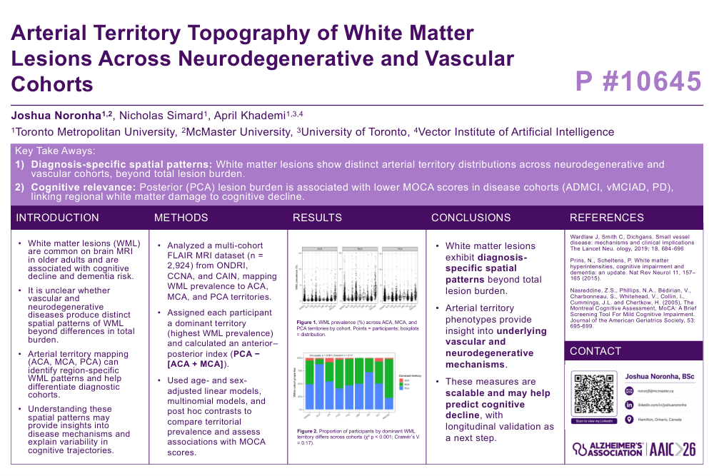
WML prevalence showed significant diagnostic-specific spatial differences across all arterial territories after adjusting for age and sex (all p < 0.001). Higher territorial prevalence (Figure 1) was found in the vascular MCI/Alzheimer’s disease (vMCIAD) cohort compared to cognitively normal (CN) participants. Spatial patterning also varied by diagnosi s (Figure 2) (χ² p < 0.001; Cramér’s V = 0.17), highlighting an MCA dominant phenotype in vMCIAD and cerebrovascular disease (CVD) patients (75% and 55%, respectively), and a PCA dominant phenotype in Parkinson’s Disease (PD) (72%) (multinomial all p < 0.001). An anterior-posterior metric further separated groups, with vMCIAD showing the strongest anterior/middle predominance (p < 0.001). The relationship between PCA prevalence and MoCA scores was diagnosis-dependent (p = 0.008), with significant negative associations in Alzheimer’s Disease MCI (ADMCI) and Frontotemporal Dementia (FTD) cohorts after multiple testing corrections (both FDR < 0.001).
Conclusion
Arterial territory mapping suggests diagnosis-specific spatial signatures that extend beyond total burden. These phenotypes may reflect distinct vascular vs neurodegenerative mechanisms and support disease characterization and stratification. Longitudinal work should test whether spatial signatures improve prediction of cognitive and clinical outcomes.
Comments
Email is required but is not shown publicly.
Loading comments...