4,068 posters, 219 videos, 101 topics, 69 sessions, 14,365 authors, 6,128 institutions
ePostersLive by SciGen Technologies S.A. All rights reserved.
Alzheimer's Association International Conference 2026
July 12-15, 2026 | London, United Kingdom, and Online

11934
Structural Signatures Of Epilepsy In Alzheimer’S Disease And Non-alzheimer Dementias: A Multicenter Exploratory Study
Poster Presenter
Authors
Part of Topic
Biomarkers -> Neuroimaging -> Disease progression measures
Here is the exact text currently on the poster, panel by panel, copied verbatim from the file. Nothing changed.
TITLE
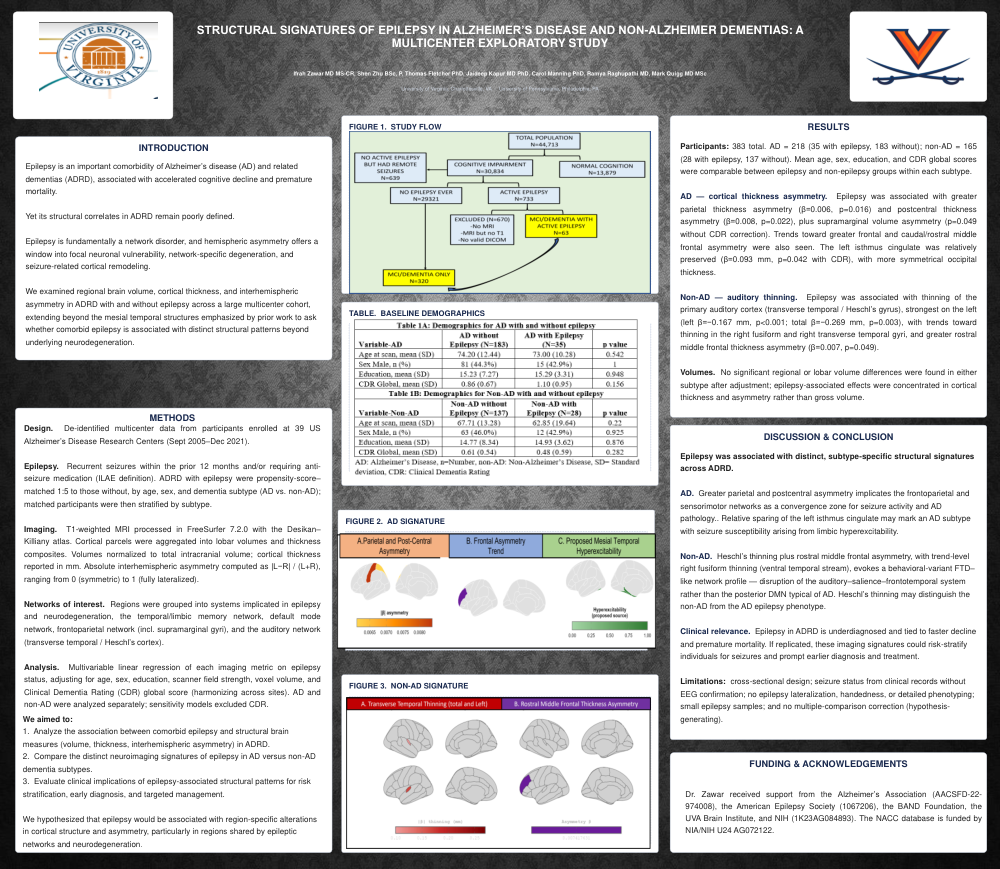
Structural Signatures of Epilepsy in Alzheimer's Disease and Non-Alzheimer Dementias: A Multicenter Exploratory Study
AUTHORS
Ifrah Zawar MD MS-CR, Shen Zhu BSc, P. Thomas Fletcher PhD, Jaideep Kapur MD PhD, Carol Manning PhD, Ramya Raghupathi MD, Mark Quigg MD MSc
INTRODUCTION
Epilepsy is an important comorbidity of Alzheimer's disease (AD) and related dementias (ADRD), associated with accelerated cognitive decline and premature mortality. Yet its structural correlates in ADRD remain poorly defined.
Epilepsy is fundamentally a network disorder, and hemispheric asymmetry offers a window into focal neuronal vulnerability, network-specific degeneration, and seizure-related cortical remodeling. We examined regional brain volume, cortical thickness, and interhemispheric asymmetry in ADRD with and without epilepsy across a large multicenter cohort, extending beyond the mesial temporal structures emphasized by prior work to ask whether comorbid epilepsy is associated with distinct structural patterns beyond underlying neurodegeneration.
METHODS
Design. De-identified multicenter data from participants enrolled at 39 US Alzheimer's Disease Research Centers (Sept 2005–Dec 2021).
Epilepsy. Recurrent seizures within the prior 12 months and/or requiring anti-seizure medication (ILAE definition). ADRD with epilepsy were propensity-score–matched 1:5 to those without, by age, sex, and dementia subtype (AD vs. non-AD); matched participants were then stratified by subtype.
Imaging. T1-weighted MRI processed in FreeSurfer 7.2.0 with the Desikan–Killiany atlas. Cortical parcels were aggregated into lobar volumes and thickness composites. Volumes normalized to total intracranial volume; cortical thickness reported in mm. Absolute interhemispheric asymmetry computed as |L−R| / (L+R), ranging from 0 (symmetric) to 1 (fully lateralized).
Networks of interest. Regions were grouped into systems implicated in epilepsy and neurodegeneration — the temporal/limbic memory network, default mode network, frontoparietal network (incl. supramarginal gyri), and the auditory network (transverse temporal / Heschl's cortex).
Analysis. Multivariable linear regression of each imaging metric on epilepsy status, adjusting for age, sex, education, scanner field strength, voxel volume, and CDR global score (harmonizing across sites). AD and non-AD analyzed separately; sensitivity models excluded CDR.
RATIONALE & OBJECTIVES
We aimed to:
- Analyze the association between comorbid epilepsy and structural brain measures (volume, thickness, interhemispheric asymmetry) in ADRD.
- Compare the distinct neuroimaging signatures of epilepsy in AD versus non-AD dementia subtypes.
- Evaluate clinical implications of epilepsy-associated structural patterns for risk stratification, early diagnosis, and targeted management.
We hypothesized that epilepsy would be associated with region-specific alterations in cortical structure and asymmetry, particularly in regions shared by epileptic networks and neurodegeneration.
RESULTS
Participants. 383 total — AD = 218 (35 with epilepsy, 183 without); non-AD = 165 (28 with epilepsy, 137 without). Mean age, sex, education, and CDR global scores were comparable between epilepsy and non-epilepsy groups within each subtype.
AD — cortical thickness asymmetry. Epilepsy was associated with greater parietal thickness asymmetry (β=0.006, p=0.016) and postcentral thickness asymmetry (β=0.008, p=0.022), plus supramarginal volume asymmetry (p=0.049 without CDR, p=0.059 with CDR). Trends toward greater frontal and rostral/caudal middle frontal asymmetry did not reach significance. In CDR-adjusted models, the left isthmus cingulate was relatively preserved (β=0.093 mm, p=0.042) and occipital thickness more symmetrical (β=−0.006, p=0.044) — marginal, specification-dependent findings to interpret with caution.
Non-AD — auditory thinning. Epilepsy was associated with thinning of the primary auditory cortex (transverse temporal / Heschl's gyrus), strongest on the left (left β=−0.167 mm, p<0.001; total β=−0.269 mm, p=0.003), with trends toward thinning in the right fusiform and right transverse temporal gyri, and greater rostral middle frontal thickness asymmetry (β=0.007, p=0.049).
Volumes. No significant regional or lobar volume differences were found in either subtype after adjustment; epilepsy-associated effects were concentrated in cortical thickness and asymmetry rather than gross volume.
DISCUSSION & CONCLUSION
Epilepsy was associated with distinct, subtype-specific structural signatures across ADRD.
AD. Greater parietal and postcentral asymmetry implicates the frontoparietal and sensorimotor networks as a convergence zone for seizure activity and AD pathology. Relative sparing of the left isthmus cingulate may mark an AD subtype with seizure susceptibility arising from limbic hyperexcitability.
Non-AD. Heschl's thinning plus rostral middle frontal asymmetry, with trend-level right fusiform thinning (ventral temporal stream), evokes a behavioral-variant FTD–like network profile — disruption of the auditory–salience–frontotemporal system rather than the posterior DMN typical of AD. Heschl's thinning may distinguish the non-AD from the AD epilepsy phenotype.
Clinical relevance. Epilepsy in ADRD is underdiagnosed and tied to faster decline and premature mortality. If replicated, these imaging signatures could risk-stratify individuals for seizures and prompt earlier diagnosis and treatment.
Limitations: cross-sectional design; seizure status from clinical records without EEG confirmation; no epilepsy lateralization, handedness, or detailed phenotyping; small epilepsy samples; and no multiple-comparison correction (hypothesis-generating).
FUNDING & ACKNOWLEDGEMENTS
Dr. Zawar received support from the Alzheimer's Association (AACSFD-22-974008), the American Epilepsy Society (1067206), the BAND Foundation, the UVA Brain Institute, and NIH (1K23AG084893). The NACC database is funded by NIA/NIH U24 AG072122.
Comments
Email is required but is not shown publicly.
Loading comments...