4,068 posters, 219 videos, 101 topics, 69 sessions, 14,365 authors, 6,128 institutions
ePostersLive by SciGen Technologies S.A. All rights reserved.
Alzheimer's Association International Conference 2026
July 12-15, 2026 | London, United Kingdom, and Online

12884
Retinal Microvascular Changes During Lecanemab Therapy In Alzheimer’S Disease: A Longitudinal Real-world Study
Part of Topic
Biomarkers -> Neuroimaging -> Evaluating treatments
INTRODUCTION: Following the approval of lecanemab for the treatment of Alzheimer’s disease (AD), there is a critical need to characterize its safety profile and associated biomarker changes in real-world clinical settings. This study evaluates longitudinal alterations in retinal microvasculature as a potential surrogate marker of cerebral microvascular changes during therapy.
METHODS: In this prospective observational study, 78 patients with AD receiving lecanemab underwent serial 3T magnetic resonance imaging (MRI) for monitoring amyloid-related imaging abnormalities (ARIA) and retinal imaging using color fundus photography (CFP). Retinal microvascular complexity was quantified using fractal dimension analysis at baseline, 6 months, and 12 months.
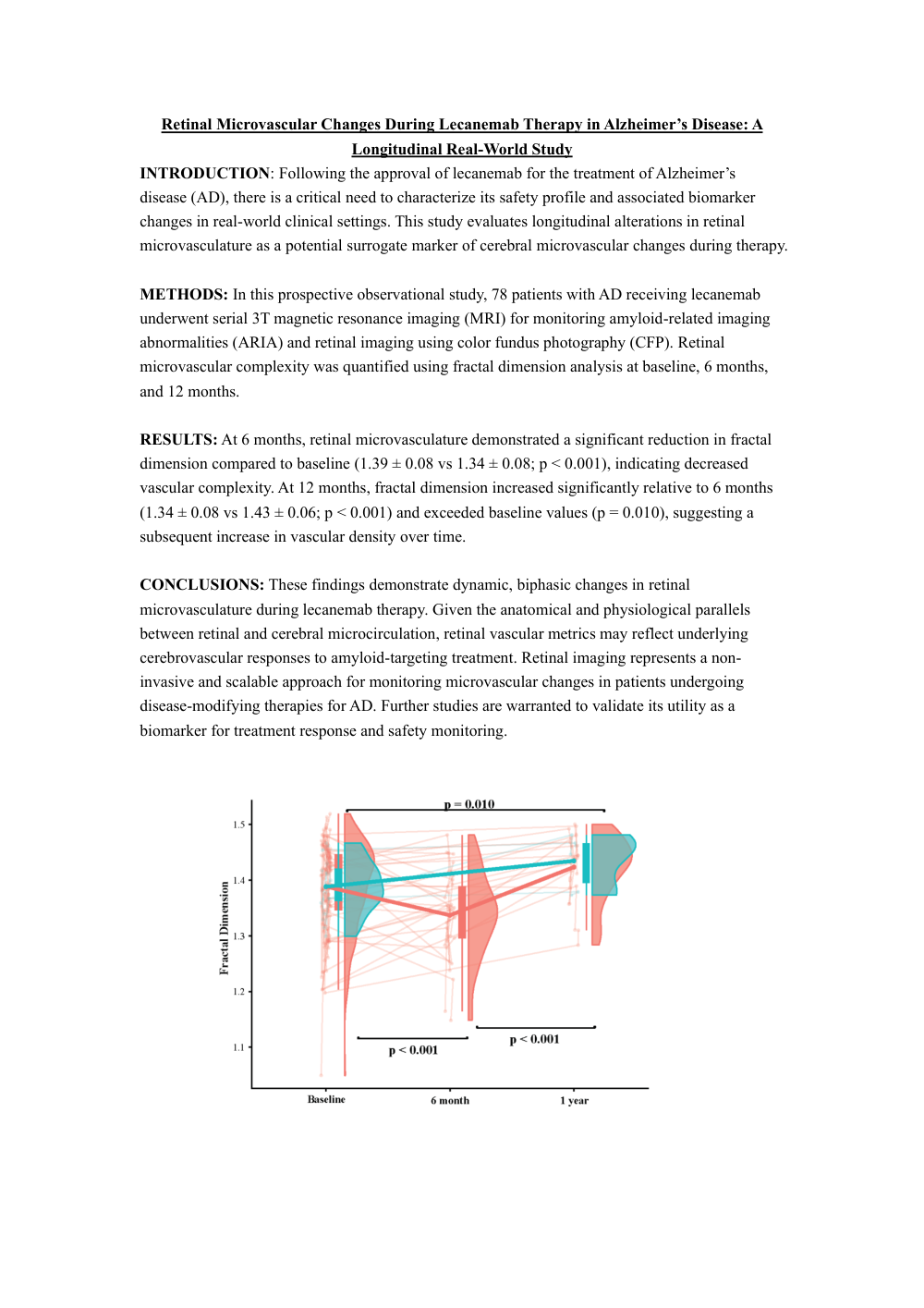
RESULTS: At 6 months, retinal microvasculature demonstrated a significant reduction in fractal dimension compared to baseline (1.39 ± 0.08 vs 1.34 ± 0.08; p < 0.001), indicating decreased vascular complexity. At 12 months, fractal dimension increased significantly relative to 6 months (1.34 ± 0.08 vs 1.43 ± 0.06; p < 0.001) and exceeded baseline values (p = 0.010), suggesting a subsequent increase in vascular density over time.
CONCLUSIONS: These findings demonstrate dynamic, biphasic changes in retinal microvasculature during lecanemab therapy. Given the anatomical and physiological parallels between retinal and cerebral microcirculation, retinal vascular metrics may reflect underlying cerebrovascular responses to amyloid-targeting treatment. Retinal imaging represents a non-invasive and scalable approach for monitoring microvascular changes in patients undergoing disease-modifying therapies for AD. Further studies are warranted to validate its utility as a biomarker for treatment response and safety monitoring.
Comments
Email is required but is not shown publicly.
Loading comments...