4,068 posters, 219 videos, 101 topics, 69 sessions, 14,365 authors, 6,128 institutions
ePostersLive by SciGen Technologies S.A. All rights reserved.
Alzheimer's Association International Conference 2026
July 12-15, 2026 | London, United Kingdom, and Online

13342
Predicting Atrophy Patterns In Frontotemporal Dementia Using Network Spreading Models And Neurotransmitter Receptor Densities
Poster Presenter
Authors
Quinten Kwok, Sonja Soskic, Elinor Thompson, Alec Sargood, Srijan Konwar, Martina Bocchetta, Daniel C. Alexander
Affiliations
UCL Queen Square Institute of Neurology, University College London, London, United Kingdom, UCL Hawkes Institute and Department of Medical Physics and Biomedical Engineering, University College London, London, United Kingdom, UCL Hawkes Institute and Department of Computer Science, University College London, London, United Kingdom, Centre for Cognitive and Clinical Neuroscience, Department of Psychology, College of Health, Medicine and Life Sciences, Brunel University of London, London, United Kingdom, Centre for Cognitive and Clinical Neuroscience, Department of Psychology, College of Health, Medicine and Life Sciences, Brunel University of London, London, United Kingdom & Dementia Research Centre, Department of Neurodegenerative Disease, UCL Queen Square Institute of Neurology, University College London, London, United Kingdom
Part of Topic
Biomarkers -> Neuroimaging -> Disease progression measures
Background
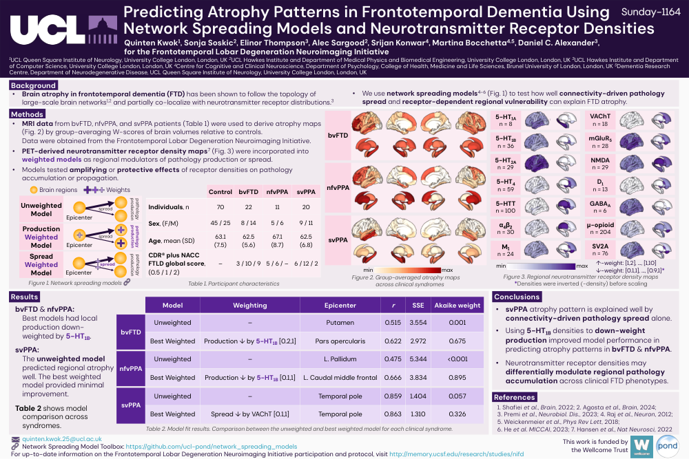
Brain atrophy in frontotemporal dementia (FTD) has been shown to follow the topology of large-scale brain networks1,2 and partially co-localize with neurotransmitter receptor distributions.3 We use network spreading models4-6 (Fig. 1) to test how well connectivity-driven pathology spread and receptor-dependent regional vulnerability can explain FTD atrophy.
Methods
MRI data from bvFTD, nfvPPA, and svPPA patients (Table 1) were used to derive atrophy maps (Fig. 2) by group-averaging W-scores of brain volumes relative to controls.
Data were obtained from the Frontotemporal Lobar Degeneration Neuroimaging Initiative.
PET-derived neurotransmitter receptor density maps7 (Fig. 3) were incorporated into weighted models as regional modulators of pathology production or spread.
Models tested amplifying or protective effects of receptor densities on pathology accumulation or propagation.
Results
bvFTD & nfvPPA: Best models had local production down-weighted by 5-HT1B.
svPPA: The unweighted model predicted regional atrophy well. The best weighted model provided minimal improvement.
Table 2 shows model comparison across syndromes.
Conclusions
svPPA atrophy pattern is explained well by connectivity-driven pathology spread alone.
Using 5-HT1B densities to down-weight production improved model performance in predicting atrophy patterns in bvFTD & nfvPPA.
Neurotransmitter receptor densities may differentially modulate regional pathology accumulation across clinical FTD phenotypes.
References
1. Shafiei et al., Brain, 2022; 2. Agosta et al., Brain, 2024; 3. Premi et al., Neurobiol. Dis., 2023; 4. Raj et al., Neuron, 2012; 5. Weickenmeier et al., Phys Rev Lett, 2018; 6. He et al. MICCAI, 2023; 7. Hansen et al., Nat Neurosci, 2022
Comments
Email is required but is not shown publicly.
Loading comments...