4,068 posters, 219 videos, 101 topics, 69 sessions, 14,365 authors, 6,128 institutions
ePostersLive by SciGen Technologies S.A. All rights reserved.
Alzheimer's Association International Conference 2026
July 12-15, 2026 | London, United Kingdom, and Online

7259
Plasma Biomarker Associations With Hippocampal Atrophy
Poster Presenter
Part of Topic
Plasma biomarker associations with hippocampal atrophy
Background: Alzheimer’s disease (AD) is characterized by amyloid-β plaques, tau neurofibrillary tangles, and neurodegeneration. Despite advances in amyloid and tau biomarkers, reliable biofluid markers of neurodegeneration remain limited. Neurofilament light chain (NfL), the most widely used marker, lacks AD specificity, reflects general axonal injury, and is influenced by both central and peripheral sources.
Objective: To identify plasma biomarkers that more specifically reflect neurodegeneration by associations with structural brain changes in AD and progression to clinical decline.
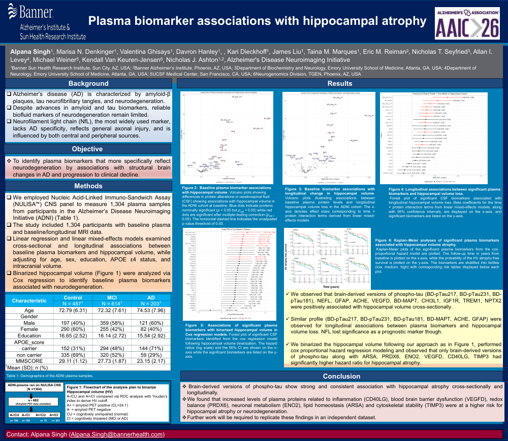
Methods: We employed Nucleic Acid-Linked Immuno-Sandwich Assay (NULISA) CNS panel to measure 1,304 plasma samples from participants in the Alzheimer’s Disease Neuroimaging Initiative (ADNI) (Table 1). The study included 1,304 participants with baseline plasma and baseline/longitudinal MRI data. Linear regression and linear mixed-effects models examined cross-sectional and longitudinal associations between baseline plasma biomarkers and hippocampal volume, while adjusting for age, sex, education, APOE ε4 status, and intracranial volume. Binarized hippocampal volume (Figure 1) were analyzed via Cox regression to identify baseline plasma biomarkers associated with neurodegeneration.
Results: We observed that brain-derived versions of phospho-tau (BD-pTau217, BD-pTau231, BD-pTau181), NEFL, GFAP, ACHE, VEGFD, BD-MAPT, CHI3L1, IGF1R, TREM1, NPTX2 were positively associated with hippocampal volume cross-sectionally. Similar profile (BD-pTau217, BD-pTau231, BD-pTau181, BD-MAPT, ACHE, GFAP) were observed for longitudinal associations between plasma biomarkers and hippocampal volume loss. NFL lost significance as a prognostic marker though. We binarized the hippocampal volume following our approach as in Figure 1, performed cox proportional hazard regression modeling and observed that only brain-derived versions of phospho-tau along with ARSA, PRDX6, ENO2, VEGFD, CD40LG, TIMP3 had significantly higher hazard ratio for hippocampal atrophy.
Conclusion: Brain-derived versions of phospho-tau show strong and consistent association with hippocampal atrophy cross-sectionally and longitudinally. We found that increased levels of plasma proteins related to inflammation (CD40LG), blood brain barrier dysfunction (VEGFD), redox balance (PRDX6), neuronal metabolism (ENO2), lipid homeostasis (ARSA) and cytoskeletal stability (TIMP3) were at a higher risk for hippocampal atrophy or neurodegeneration. Further work will be required to replicate these findings in an independent dataset.
Comments
Email is required but is not shown publicly.
Loading comments...