4,068 posters, 219 videos, 101 topics, 69 sessions, 14,365 authors, 6,128 institutions
ePostersLive by SciGen Technologies S.A. All rights reserved.
Alzheimer's Association International Conference 2026
July 12-15, 2026 | London, United Kingdom, and Online

13884
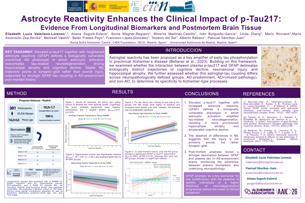
Astrocyte Reactivity Enhances The Clinical Impact Of p-Tau217: Evidence From Longitudinal Biomarkers And PostMortem Brain Tissue
Poster Presenter
Authors
Elizabeth Lucía Valeriano-Lorenzo, Sonia Wagner-Reguero, Minerva Martínez-Castillo, Iván Burgueño-García, Linda Zhang, Mario Ricciardi, María Ascensión Zea-Sevilla, Meritxell Valentí, Belén Frades-Payo, Francisco López-González, Teodoro Del Ser, Alberto Rábano, Aitana Sogorb-Esteve, Pascual Sánchez-Juan
Affiliations
Part of Topic
Biomarkers -> Biomarkers (non-neuroimaging) -> Longitudinal
Abstract:
Background:
Astroglial reactivity has been proposed as a key amplifier of early tau phosphorylation inpreclinical Alzheimer’s disease (Bellaver et al., 2023). Building on this framework, we examined whetherthe interaction between plasma p‑tau217 and GFAP delineates biologically distinct trajectories ofcognitive decline, neuroaxonal injury, and hippocampal atrophy. We further assessed whether thisastroglial-tau coupling differs across neuropathologically defined groups, AD‑predominant, AD+(mixed pathology), and non‑AD, to determine its specificity to Alzheimer‑type processes.
Methods:
We studied 381 cognitively unimpaired older adults at baseline (mean age 74.2 ± 3.5 years;61.2% women) with repeated plasma biomarkers, structural MRI, and ≥3 cognitive assessments over upto 10 years. Plasma p‑tau217 was quantified using LC-MS/MS, and GFAP and NfL levels using SIMOA(Quanterix SR‑X). Astroglial status (Ast+ vs. Ast-) was derived using a data‑driven cutoff based on theYouden index from ROC analyses. Participants were classified into four groups based on p‑tau217status and astrocyte reactivity (Table 1). Longitudinal change was modeled with: a GAMM for globalcognition, a GAM for plasma NfL, and a GAMM for hippocampal volume (ICV‑adjusted), all includingsmooth time‑by‑group terms and standard covariates. We analyzed a second cohort of 100 braindonors using partial correlations adjusted for age at extraction, estimated symptom‑onset age, sex, andThal stage.
Results:
Across all outcomes, the Ast+/p‑tau+ group showed the most adverse profile. Cognitively, thisgroup exhibited significantly lower baseline performance and steeper decline (βGroup4=0.41, p<0.001) (Figure 1).NfL levels were higher at baseline and increased more rapidly over time however the estimate was notstatistically significant (Figure 3). Hippocampal volume was significantly reduced (βGroup4= -76.1 mm³, p= 0.031) and declined faster than in all other groups (Figure2). In post-mortem tissue, only the AD group showed a significant GFAP-p-tau217 association (βAD=0.0083, pFDR=0.010), whereas AD+ and non-ADgroups showed no significant effects (Figure4).
Conclusion:
The interaction between p‑tau217 and astrocyte reactivity identifies a biologicallyvulnerable phenotype characterized by accelerated cognitive decline, heightened neuroaxonaldegeneration, pronounced hippocampal atrophy, and stronger GFAP-tau coupling in post‑mortemtissue. These findings position GFAP as a key amplifier of early tau‑related neurodegeneration and apromising biomarker for risk stratification in preclinical AD.
Comments
Email is required but is not shown publicly.
Loading comments...