4,068 posters, 219 videos, 101 topics, 69 sessions, 14,365 authors, 6,128 institutions
ePostersLive by SciGen Technologies S.A. All rights reserved.
Alzheimer's Association International Conference 2026
July 12-15, 2026 | London, United Kingdom, and Online

9725
Age-dependent Changes In Brain Rhythms And Visual Responses In A Humanised Mouse Model Of Tauopathy
Poster Presenter
Authors
Fabio Ribeiro Rodrigues, Joanna Holeniewska, Sara de Leo, Aman Saleem, Samuel Solomon
Affiliations
Part of Topic
Introduction
Dementia is commonly associated with malfunction of Tau protein, resulting in hyperphosphorylated aggregates in neurons. This causes synaptic dysfunction, neuronal hypoactivity, and neurodegeneration. In multiple mouse models, tauopathy results in increases in delta range activity, and reductions in theta and gamma power during resting state.
Newer mouse models of tauopathy have been generated by humanising the mouse MAPT gene, with or without familial mutations of frontotemporal dementia. Unlike previous overexpression models, humanised Tau (hTau) animals with mutations develop pathology at more advanced ages, providing a unique opportunity to evaluate the early impact of pathology on brain activity and behaviour.
Here we evaluate how pathology progressively affects brain function in a humanised mouse model of frontotemporal dementia, at multiple ages
Methods
We used humanised Tau (hTau) animals as Controls, and hTau animals carrying mutations associated with familial forms of frontotemporal dementia (Tau+). These animals have increased levels of phosphorylated Tau from about 15 months of age.
Animals were implanted with local field potential (LFP) electrodes in prefrontal cortex (PFC), CA1, left and right visual cortex (V1) and ectorhinal cortex (ECT), at 8-11 months, and recorded until 21 months of age. Speed of locomotion and pupil size were monitored throughout recording sessions. Data is presented as mean±sem, and individual animal values are represented by circles. All comparisons were made using a repeated measured mixed ANOVA, and post-hoc tests were Bonferroni corrected (*, p<0.05; **, p<0.01; ***, p<0.001).
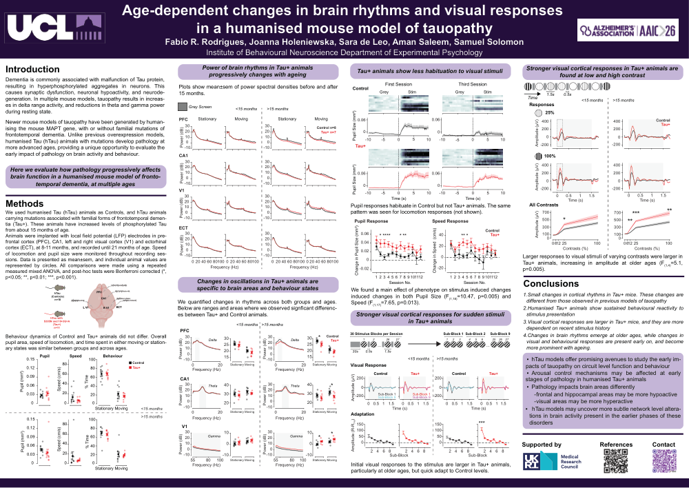
Behaviour dynamics of Control and Tau+ animals did not differ. Overall pupil area, speed of locomotion, and time spent in either moving or stationary states was similar between groups and across ages.
Power of brain rhythms in Tau+ animals progressively changes with ageing
Plots show mean±sem of power spectral densities before and after 15 months.
Changes in oscillations in Tau+ animals are specific to brain areas and behaviour states
We quantified changes in rhythms across both groups and ages. Below are ranges and areas where we observed signficant differences between Tau+ and Control animals.
Tau+ animals show less habituation to visual stimuli
We found a main effect of phenotype on stimulus induced changes induced changes in both Pupil Size (F(1,18)=10.47, p=0.005) and Speed (F(1,17)=7.65, p=0.013).
Stronger visual cortical responses for sudden stimuli in Tau+ animals
Initial visual responses to the stimulus are larger in Tau+ animals, particularly at older ages, but quick adapt to Control levels.
Stronger visual cortical responses in Tau+ animals are found at low and high contrast
Larger responses to visual stimuli of varying contrasts were larger in Tau+ animals, increasing in amplitude at older ages (F(1,4)=5.1, p=0.005).
Conclusions
1. Small changes in cortical rhythms in Tau+ mice. These changes are different from those observed in previous models of tauopathy
2. Humanised Tau+ animals show sustained behavioural reactivity to stimulus presentation
3. Visual cortical responses are larger in Tau+ mice, and they are more dependent on recent stimulus history
4. Changes in brain rhythms emerge at older ages, while changes in visual and behavioural responses are present early on, and become more prominent with ageing.
hTau models offer promising avenues to study the early impacts of tauopathy on circuit level function and behaviour.
Arousal control mechanisms may be affected at early stages of pathology in humanised Tau+ animals.
Pathology impacts brain areas differently: frontal and hippocampal areas may be more hypoactive, visual areas may be more hyperactive.hTau models may uncover more subtle network level alterations in brain activity present in the earlier phases of these disorders
Comments
Email is required but is not shown publicly.
Loading comments...